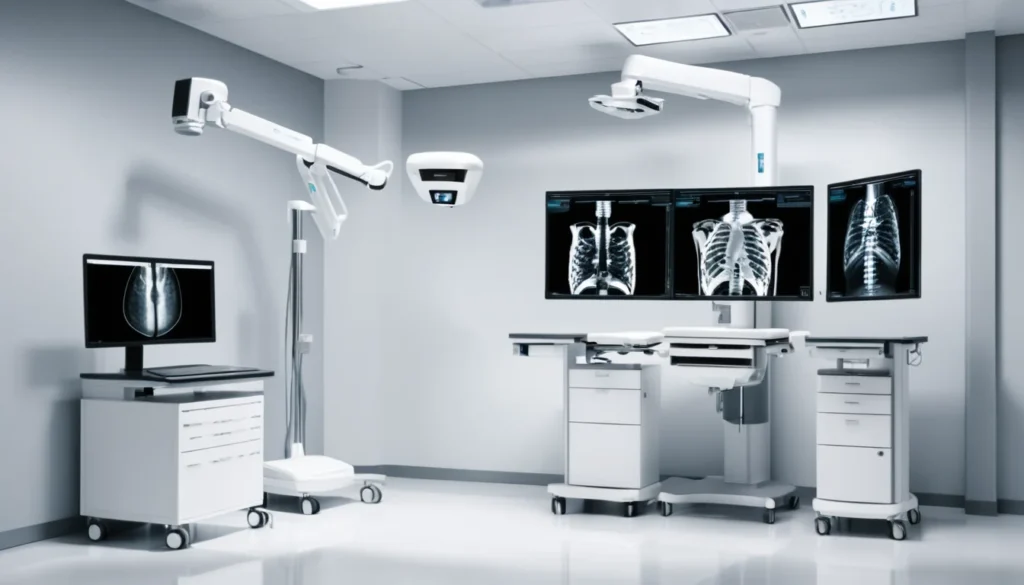
X-rays are one of the most common medical tools used across Canada today. They help doctors see inside your body without making a single cut. In this article, we explain how x-rays work, how they are used to diagnose and treat illness, and what you should know before your next scan.
What Are X-Rays?
X-rays are a form of electromagnetic radiation. Like visible light, they travel in waves — but their wavelengths are far too short for the human eye to see. In fact, x-ray wavelengths are measured in units called angstroms, which are incredibly tiny.
Because of their short wavelength, x-rays carry enough energy to pass through soft tissue, muscle, and organs. This is what makes them so useful in medicine. However, denser materials like bone absorb more radiation, which is why bones appear white on an x-ray image.
X-rays belong to the same family of waves as radio waves, microwaves, and ultraviolet light. The key difference is energy level. X-rays have much higher energy, which gives them their unique ability to penetrate living tissue. The World Health Organization explains more about ionizing radiation and its effects on health.
How X-Rays Are Used in Diagnosis
In Canada, x-rays are a core part of diagnostic medicine. Your family doctor or a specialist may order an x-ray to get a clearer picture of what is happening inside your body. Hospitals, walk-in clinics, and imaging centres across every province offer this service.
X-ray imaging falls under a branch of medicine called radiology. A radiologist is a doctor who specialises in reading and interpreting medical images. After your scan, the radiologist sends a report to your referring doctor, who then discusses the results with you.
Common Reasons for a Diagnostic X-Ray
Doctors use x-rays to diagnose a wide range of conditions. Here are some of the most common reasons you might be sent for one:
Suspected broken or fractured bones
Chest infections such as pneumonia
Joint problems like arthritis
Dental issues, including cavities and jaw problems
Swallowed objects, especially in children
Lung conditions, including monitoring for certain cancers
In most provinces, a referral from your family doctor or walk-in clinic is needed to access imaging services covered by your provincial health plan. For example, in Ontario this falls under OHIP, while in British Columbia it is covered by MSP. Always check with your provider to understand your coverage.
Conventional X-Ray vs. CT Scan
A standard x-ray produces a flat, two-dimensional image. It is quick, widely available, and uses a relatively low dose of radiation. This makes it ideal for a first look at bones, lungs, and the chest area.
A CT scan — also called computed tomography or a CAT scan — is a more advanced form of x-ray imaging. It takes many images from different angles and uses a computer to build a detailed, three-dimensional picture. CT scans are often used when doctors need more precise information about organs, blood vessels, or soft tissue. However, because CT scans involve more radiation than a standard x-ray, doctors only order them when the benefit clearly outweighs the risk.
Both types of x-ray imaging are widely available through Canadian hospitals and diagnostic imaging centres. Your doctor will choose the right type based on your symptoms and medical history. The Mayo Clinic offers a helpful overview of what to expect during an x-ray procedure.
How X-Rays Are Used in Treatment
Beyond diagnosis, x-rays also play a role in treating certain medical conditions. This is known as radiation therapy, or radiotherapy. It is most commonly used in the treatment of cancer.
External beam radiation therapy directs focused beams of radiation at a tumour. The goal is to damage the DNA of cancer cells so they can no longer grow or divide. Healthy tissue around the tumour can sometimes be affected too, which is why treatment is carefully planned and targeted.
X-Rays vs. More Powerful Radiation in Cancer Treatment
Standard x-rays have a relatively low ability to ionise — meaning they are not always the most powerful tool for cancer treatment on their own. As a result, cancer treatment centres often use higher-energy radiation sources.
One example is gamma radiation, which is produced during cobalt therapy. Cobalt-60 machines release gamma rays that are more energetic than standard x-rays. This makes them more effective at targeting deep tumours. Today, many cancer centres have moved toward even more advanced technologies such as linear accelerators, which produce high-energy x-rays that can be precisely aimed. Canadian cancer patients can access these treatments through provincial cancer agencies such as Cancer Care Ontario or BC Cancer.
Furthermore, newer techniques like intensity-modulated radiation therapy (IMRT) allow doctors to shape the radiation beam to match the exact shape of a tumour. This helps protect nearby healthy tissue. Radiation oncologists — doctors who specialise in cancer treatment using radiation — oversee these programmes.
Understanding Radiation Safety
It is natural to feel concerned about radiation when your doctor orders an x-ray. However, the doses used in medical imaging are generally very low. For most people, the benefit of getting an accurate diagnosis far outweighs the small risk from radiation exposure.
To put it in perspective, a single chest x-ray exposes you to roughly the same amount of radiation you would receive from a few hours of natural background radiation — the kind that surrounds us every day from soil, rocks, and even the air. A CT scan involves more exposure, but it is still considered safe when used appropriately.
Who Should Take Extra Precautions?
Certain groups of people need extra care when it comes to x-ray exposure. These include:
Pregnant women: X-rays are generally avoided during pregnancy unless absolutely necessary, especially during the first trimester. Always tell your doctor or technologist if you are pregnant or think you might be.
Children: Young children are more sensitive to radiation because their cells are still developing. Doctors follow strict guidelines to use the lowest possible dose.
People with frequent imaging needs: If you require many x-rays over time due to a chronic condition, your doctor will track your cumulative exposure and adjust imaging plans accordingly.
In Canada, medical radiation technologists (MRTs) are regulated health professionals. They are trained to use the lowest effective dose of radiation for every scan. Protective equipment such as lead aprons is available and used when appropriate. Health Canada provides guidance on understanding radiation in everyday life.
The ALARA Principle
Medical professionals in Canada follow a safety standard known as ALARA — As Low As Reasonably Achievable. This means they always use the minimum amount of radiation needed to get a clear, useful image. Your safety is always the top priority during any imaging procedure.
What to Expect During an X-Ray
Getting an x-ray is a straightforward and painless procedure. Most standard x-rays take only a few minutes from start to finish. Here is what typically happens:
You may be asked to remove jewellery, eyeglasses, or clothing with metal fasteners in the area being imaged.
You will be positioned by the technologist — either standing, sitting, or lying down — depending on the body part being examined.
You will need to stay very still for a moment while the image is taken. Movement can blur the picture.
In some cases, you may need images taken from more than one angle.
The procedure itself is completely painless. You will not feel the x-rays passing through your body.
After your x-ray, you can go about your normal day right away. There is no recovery time needed. Results are usually sent to your doctor within a day or two, though this can vary by province and facility.
When to See a Doctor
If your family doctor or a walk-in clinic physician has recommended an x-ray, it is important to follow through with that referral. Early imaging can catch problems before they become more serious.
You should speak with your doctor if you have concerns about radiation exposure, if you are pregnant, or if you have had many x-rays in the past. Your doctor can help you weigh the benefits against any potential risks. Do not skip an x-ray that your doctor has recommended out of fear — the information it provides is usually far more valuable than the minimal risk involved.
If you do not have a family doctor, a walk-in clinic can refer you for many types of imaging. You can also contact Health811 (formerly Telehealth Ontario) or your province’s equivalent health line for guidance on next steps. As always, consult a qualified healthcare provider before making any decisions about your health or treatment.
Frequently Asked Questions About X-Rays
Are x-rays safe for adults?
For most adults, x-rays are considered very safe. The dose of radiation used in a standard medical x-ray is very low — similar to natural background radiation you encounter every day. Your doctor will only recommend an x-ray when the benefit of diagnosis clearly outweighs any minimal risk.
Can x-rays detect cancer?
X-rays can detect some signs of cancer, such as unusual masses or shadows in the lungs or bones. However, they are not always the most detailed imaging tool. Doctors often follow up with a CT scan, MRI, or biopsy for a more complete picture if cancer is suspected.
What is the difference between an x-ray and a CT scan?
A standard x-ray produces a flat, two-dimensional image and uses a low dose of radiation. A CT scan uses multiple x-ray images taken from different angles to create a detailed three-dimensional view. CT scans provide more information but involve a higher dose of radiation, so they are used only when necessary.
Are x-rays covered by provincial health plans in Canada?
In most provinces, medically necessary x-rays ordered by a licensed physician are covered under provincial health plans such as OHIP in Ontario or MSP in British Columbia. Coverage details can vary, so it is best to confirm with your doctor and your provincial health authority before your appointment.
Is it safe to have an x-ray during pregnancy?
Doctors generally avoid x-rays during pregnancy unless absolutely necessary, particularly during the first trimester when the baby is developing rapidly. Always tell your doctor or the imaging technologist if you are pregnant or suspect you might be, so they can take appropriate precautions or consider alternative imaging.
How long does it take to get x-ray results in Canada?
After your x-ray, a radiologist will review the images and send a report to your referring doctor. This process usually takes one to two business days, though urgent cases may be reviewed more quickly. Your family doctor or walk-in clinic physician will contact you to discuss the results.
Key Takeaways
X-rays are a safe and widely used form of electromagnetic radiation in Canadian medicine.
They are used for diagnosis through conventional imaging and CT scans, helping doctors see inside the body without surgery.
X-rays also play a role in cancer treatment through radiation therapy, though higher-energy radiation is often preferred for deep tumours.
The ALARA principle ensures that radiation doses are kept as low as possible in all medical settings across Canada.
Certain groups — including pregnant women and children — should take extra precautions and always inform their healthcare provider.
Most medically necessary x-rays are covered by provincial health plans. Check with your provider for details.
Always consult your family doctor or a walk-in clinic if you have questions or concerns about x-ray imaging.