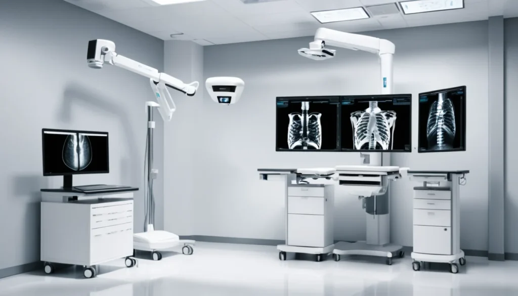
An x-ray is one of the most common medical imaging tests used in Canada today. It uses a small amount of radiation to create a picture of the inside of your body, helping doctors spot broken bones, infections, and other health concerns. This guide explains how x-rays work, why your doctor might order one, and exactly what happens during the procedure.
What Is an X-Ray?
An x-ray is a type of medical image taken by passing a beam of radiation through the body onto a film or digital sensor. Dense tissues like bone block most of the radiation and show up white on the image. Softer tissues, such as muscles and organs, allow more radiation to pass through and appear in shades of grey.
The result is a detailed picture — called a radiograph — that lets your doctor see what is happening inside your body without any cuts or surgery. X-rays are fast, painless, and widely available across Canada, from major hospitals to smaller community health centres.
How Does an X-Ray Work?
An x-ray machine sends a focused beam of electromagnetic radiation through the part of your body being examined. Different body tissues absorb different amounts of radiation. As a result, the image that forms on the other side shows a map of those differences.
For example, a broken bone looks different from a healthy one because the fracture changes how the bone absorbs radiation. Air-filled spaces, like your lungs, absorb very little radiation and appear dark on the image. This contrast is what makes x-rays so useful for diagnosing a wide range of conditions.
Digital vs. Traditional X-Rays
Most Canadian hospitals and clinics now use digital x-ray technology instead of old-fashioned film. Digital x-rays produce clearer images and are available to your doctor almost instantly. They also use slightly less radiation than traditional film-based systems.
Furthermore, digital images can be stored electronically and shared quickly between your family doctor, a specialist, or a walk-in clinic. This makes follow-up care much faster and more efficient within Canada’s provincial health systems.
Common Reasons a Doctor Orders an X-Ray
Doctors order x-rays for many different reasons. The most common is to check for a broken or fractured bone after an injury. However, x-rays are useful for much more than broken bones.
Your doctor may order an x-ray to help diagnose or monitor any of the following conditions:
Broken or fractured bones — including hairline fractures that may not be visible otherwise
Chest infections — such as pneumonia, which can show up as cloudy patches in the lungs
Arthritis — to check for joint damage and bone changes over time
Heart and lung conditions — a chest x-ray can show an enlarged heart or fluid in the lungs
Dental problems — dentists use x-rays to spot cavities, impacted teeth, and bone loss
Swallowed objects — especially important in young children
Digestive issues — to look for blockages or abnormal gas patterns in the intestines
Certain cancers — some tumours are visible on plain x-rays, though CT scans are usually needed for more detail
In addition, x-rays are often the first step in a broader diagnostic process. If your x-ray raises questions, your doctor may refer you for more advanced imaging such as an ultrasound, MRI, or CT scan.
What to Expect During an X-Ray
Getting an x-ray is a simple and quick process. In most cases, the entire procedure takes less than 15 minutes from start to finish. You do not need to do anything special to prepare for a standard x-ray of a bone or your chest.
Before the X-Ray
Before the scan, a technologist — the health professional who operates the x-ray machine — will ask you to remove any jewellery, piercings, or clothing with metal fasteners near the area being imaged. Metal objects block radiation and can interfere with the image. You may be given a gown to wear instead.
If you are pregnant or think you might be pregnant, tell the technologist right away. While the radiation dose from a single x-ray is very low, doctors usually avoid x-raying the abdomen or pelvis during pregnancy unless it is medically necessary. Health Canada provides guidance on radiation safety in medical imaging.
During the X-Ray
The technologist will position you carefully so the x-ray captures the right area. You may need to stand, sit, or lie down depending on what part of your body is being examined. The technologist will step behind a protective screen or into a separate room to take the image.
You will be asked to stay very still — and sometimes to hold your breath for a second — while the image is taken. Movement can blur the picture. The actual exposure to radiation lasts only a fraction of a second. You will not feel anything at all during the x-ray itself.
After the X-Ray
Once the images are taken, a radiologist — a doctor who specialises in reading medical images — will study the results. They will send a written report to the doctor who ordered your x-ray. Depending on the setting, you may get your results the same day or within a few days.
Your family doctor or the clinic that sent you for the x-ray will contact you to discuss the findings. If results are urgent, you will typically hear back sooner. Do not hesitate to follow up with your doctor if you have not received your results within the expected timeframe.
Are X-Rays Safe?
X-rays do involve a small amount of ionising radiation. However, the dose used in a standard diagnostic x-ray is very low and considered safe for most people. To put it in perspective, a single chest x-ray exposes you to roughly the same amount of radiation you would receive from a few hours of natural background radiation in daily life.
That said, radiation exposure does accumulate over a lifetime. Therefore, doctors only order x-rays when there is a clear medical reason to do so. According to the Mayo Clinic, the benefit of a medically necessary x-ray almost always outweighs the small radiation risk.
Protective Measures During X-Rays
Radiology technologists are trained to use the lowest possible radiation dose to get a clear image. They also use lead shields — heavy aprons or collars — to protect parts of your body that do not need to be imaged, such as your thyroid gland or reproductive organs.
Children and pregnant individuals receive extra care and protection. In many cases, alternative imaging methods that use no radiation — such as ultrasound or MRI — are considered first for these groups.
Types of X-Ray Procedures
Not all x-ray procedures are the same. Some are straightforward, while others involve a special contrast material to make certain tissues easier to see. Here are the most common types you might encounter in Canada:
Plain x-ray (radiograph) — the standard, everyday x-ray used for bones and chest imaging
Chest x-ray — checks the lungs, heart, and ribs; one of the most frequently ordered x-rays in Canada
Dental x-ray — used by your dentist to examine teeth and jaw bones
Fluoroscopy — a real-time x-ray that shows moving images, often used to examine the digestive tract
Mammography — a specialised low-dose x-ray of breast tissue used in cancer screening programmes
Bone density scan (DEXA) — measures bone strength and screens for osteoporosis
Your doctor will choose the right type of x-ray based on your symptoms and what they need to find out. Healthline offers a helpful overview of different x-ray types and their uses.
X-Rays and the Canadian Healthcare System
In Canada, x-rays ordered by a doctor are generally covered by your provincial or territorial health plan. This means there is usually no out-of-pocket cost for a medically necessary x-ray. Coverage details can vary slightly by province, so it is worth checking with your provincial health authority if you are unsure.
Your family doctor or a walk-in clinic doctor can refer you for an x-ray. In some cases, such as an emergency department visit, x-rays may be done immediately on site. Radiology centres and hospital imaging departments across the country perform thousands of x-rays every day as a routine part of patient care.
Wait times for non-urgent x-rays are generally short — often just a day or two. If your doctor suspects something serious, they can mark the referral as urgent to ensure you are seen faster.
When to See a Doctor
You should speak with your family doctor or visit a walk-in clinic if you have symptoms that might need an x-ray. Common signs include pain or swelling after an injury, persistent chest pain or cough, or breathing difficulties that do not improve on their own.
If you have already had an x-ray and are waiting for results, contact the clinic or your doctor’s office if you have not heard back within the timeframe they gave you. Do not ignore worsening symptoms while waiting. If your symptoms become severe — such as difficulty breathing, chest pain, or signs of a serious injury — go to your nearest emergency department right away.
Always discuss your medical imaging results with a qualified healthcare provider. A radiograph is a tool to help your doctor make decisions — it is not something you should try to interpret on your own. As always, consult your doctor before drawing any conclusions about your health based on medical test results.
Frequently Asked Questions About X-Rays
Is an x-ray painful?
No, an x-ray is completely painless. You will not feel the radiation at all during the procedure. The only discomfort you might experience is from holding a certain position if you have an existing injury.
How long does an x-ray take?
A standard x-ray procedure takes about 10 to 15 minutes in total, including preparation time. The actual exposure to radiation lasts less than a second. More complex x-ray procedures, such as a barium swallow, may take longer.
Can I get an x-ray if I am pregnant?
It is important to tell your doctor and the x-ray technologist if you are pregnant or think you might be. X-rays of the chest or limbs are generally considered safe during pregnancy, but abdominal x-rays are avoided when possible. Your doctor will weigh the risks and benefits carefully before ordering any imaging during pregnancy.
Is an x-ray covered by provincial health insurance in Canada?
Yes, in most cases a medically necessary x-ray ordered by a doctor is covered under your provincial or territorial health plan at no direct cost to you. Coverage can vary slightly by province, so check with your local health authority if you have questions about your specific situation.
What is the difference between an x-ray and a CT scan?
An x-ray produces a single flat image of a body part, while a CT (computed tomography) scan takes many x-ray images from different angles and combines them into a detailed 3D picture. CT scans provide much more detail but also use a higher dose of radiation. Your doctor will choose the right type of imaging based on what they need to see.
How do I get my x-ray results in Canada?
After your x-ray, a radiologist reviews the images and sends a report to the doctor who referred you. Your family doctor or the walk-in clinic will then contact you to discuss the results, usually within a few days. If you have not heard back within the expected time, call your doctor’s office to follow up.
Key Takeaways
An x-ray is a quick, painless imaging test that uses a small amount of radiation to create pictures of the inside of your body.
X-rays are used to diagnose broken bones, chest infections, arthritis, dental problems, and many other conditions.
The radiation dose from a standard x-ray is very low and considered safe when medically necessary.
In Canada, medically necessary x-rays are generally covered by your provincial or territorial health plan.
Your family doctor or a walk-in clinic can refer you for an x-ray; results are reviewed by a radiologist and shared with your doctor.
Always consult your doctor to understand your x-ray results and what they mean for your health.