The retina is one of the most important parts of your eye. It is a thin layer of tissue that lines the inside back wall of the eyeball and makes vision possible. Without a healthy retina, your brain cannot receive or process visual information. In this article, we explain how the retina works, what can go wrong, and when to talk to your family doctor or visit a walk-in clinic.
What Is the Retina?
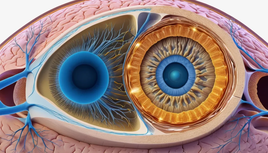
The retina is a thin, transparent membrane located at the back of your eye. Think of it like the film in an old camera — it captures light and turns it into signals your brain can understand.
The retina sits between two important layers. Its back surface touches the choroid, a blood vessel-rich layer, through a special lining called the retinal pigment epithelium. Its front surface faces the vitreous body, the clear gel that fills the inside of your eye.
Blood reaches the retina through the central retinal artery. This artery enters the eye through the optic disc — the point where the optic nerve begins. Proper blood flow is essential for keeping the retina healthy and working correctly.
How the Retina Works: Structure and Function
The retina contains several layers of specialized cells. Three of these layers work together to detect light and send visual signals to the brain.
The Three Main Cell Layers
Starting from the back of the eye and moving forward, the three key layers are:
Photoreceptor cells — These sit at the very back of the retina. They include rods and cones. Rods help you see in low light and detect movement. Cones give you sharp, colour vision in bright light.
Bipolar cells — These act as a relay station. They receive signals from the photoreceptors and pass them forward.
Ganglion cells — These sit at the front of the retina. They collect signals from the bipolar cells and send them down the optic nerve to the brain.
This three-layer system processes visual information quickly and efficiently. As a result, your brain receives a clear picture of the world around you almost instantly.
Rods vs. Cones: What Is the Difference?
Your retina contains about 120 million rod cells and 6 million cone cells. Rods are spread across the entire retina, while cones are concentrated in the centre, in an area called the macula.
The macula is responsible for your central vision — the sharp focus you use for reading, recognizing faces, and driving. When the macula is damaged, everyday tasks become much harder.
How Doctors Examine the Retina
Eye doctors use several methods to examine the retina. The most common is a fundus examination, also known as looking at the “back of the eye.” Your doctor may use eye drops to widen (dilate) your pupils first. This gives them a better view of the retina’s full surface.
Advanced Retinal Tests
In addition to a basic eye exam, specialists can use more detailed tests to study how your retina works and what it looks like:
Electroretinography (ERG) — This test measures the electrical activity of your retinal cells when exposed to light. It helps detect conditions affecting the photoreceptors.
Electrooculography (EOG) — This measures the electrical potential between the front and back of your eye. It checks how well the retinal pigment epithelium is functioning.
Ocular angiography — A dye is injected into a vein in your arm. A special camera then photographs the blood vessels in your retina. This reveals blockages, leaks, or abnormal vessel growth.
These tests are usually done by an ophthalmologist (an eye specialist). Your family doctor can refer you if there is a concern. Most provincial health plans in Canada cover medically necessary eye exams and specialist referrals. However, routine adult eye exams may not be covered in all provinces, so check with your provincial health plan for details.
Common Retinal Diseases and Conditions
Several diseases can affect the retina. Some develop slowly over time, while others happen suddenly and require urgent care. Understanding the most common ones can help you recognize warning signs early.
Retinal Detachment
Retinal detachment occurs when the retina peels away from the back of the eye. This is a medical emergency. Without prompt treatment, it can cause permanent vision loss.
It is more common in people who are nearsighted (myopic), older adults, or those with a family history of the condition. Warning signs include sudden flashes of light, a large number of new floaters, or a shadow or curtain across your vision. If you notice any of these symptoms, go to an emergency room or walk-in clinic right away.
For more information, visit Mayo Clinic’s guide to retinal detachment.
Degenerative Retinal Diseases
Degenerative retinal diseases damage the photoreceptor cells and/or the retinal pigment epithelium over time. Many of these conditions are inherited, meaning they run in families.
Two well-known examples are:
Retinitis pigmentosa — A group of genetic disorders that gradually break down the rod cells. Night blindness is often the first symptom, followed by a narrowing of peripheral (side) vision.
Tapeto-retinal degeneration — A broader term for inherited conditions that damage multiple layers of the retina over time.
Furthermore, age-related macular degeneration (AMD) is one of the leading causes of vision loss in Canadians over 50. It damages the macula and affects central vision. Health Canada recognizes AMD as a growing public health concern as Canada’s population ages.
Inflammatory Retinal Conditions
Inflammation of the retina is relatively rare. However, it can be serious. In most cases, retinal inflammation (called retinitis) is caused by inflammation spreading from the choroid layer beneath it — a condition called chorioretinitis.
One of the most common causes of chorioretinitis worldwide is toxoplasmosis, an infection caused by a parasite called Toxoplasma gondii. It can be contracted through undercooked meat or contact with infected cat feces. People with weakened immune systems are especially at risk.
Vascular Retinal Diseases
Vascular diseases affect the blood vessels that supply the retina. Two main types of blockages can occur:
Central retinal artery occlusion — The main artery supplying the retina becomes blocked. This causes sudden, painless vision loss. It is treated as a medical emergency.
Central retinal vein occlusion — The main vein draining blood from the retina becomes blocked. This leads to swelling and bleeding in the retina.
In addition, the tiny blood vessels (microcirculation) of the retina can be damaged by diabetes and high blood pressure (hypertension). This is called diabetic retinopathy or hypertensive retinopathy. Diabetic retinopathy is one of the most common causes of preventable blindness in Canada. Therefore, Canadians living with diabetes should have regular eye exams. Learn more at Healthline’s overview of diabetic retinopathy.
Retinal Tumours
Tumours affecting the retina are uncommon. However, the most important one to know about is retinoblastoma. This is a malignant (cancerous) tumour that almost exclusively affects very young children, usually under the age of five.
A key warning sign is a white or yellowish glow in the pupil, visible in photographs taken with a flash. This is sometimes called “cat’s eye reflex.” Early diagnosis greatly improves outcomes, so parents should mention any unusual pupil appearance to their child’s doctor right away.
Risk Factors for Retinal Problems
Certain factors increase your chances of developing a retinal disease. Knowing your risk can help you take action early.
Age — The risk of macular degeneration and retinal detachment increases significantly after age 50.
Diabetes — High blood sugar damages small blood vessels throughout the body, including those in the retina.
High blood pressure — Uncontrolled hypertension puts stress on retinal blood vessels.
Nearsightedness (myopia) — A higher degree of myopia stretches the retina and raises the risk of detachment.
Family history — Many retinal diseases have a genetic component. Knowing your family’s eye health history is important.
Eye injuries — Trauma to the eye can trigger retinal tears or detachment.
When to See a Doctor
You should contact your family doctor or visit a walk-in clinic if you notice any of the following warning signs related to your retina or general vision:
Sudden flashes of light in one or both eyes
A sudden increase in floaters (spots or strings drifting across your vision)
A dark shadow or curtain covering part of your visual field
Blurred or distorted central vision
Difficulty seeing in low-light conditions
A white glow in a child’s pupil in photographs
Retinal detachment and artery blockages are emergencies. Do not wait for a regular appointment if you experience sudden vision changes. Go to your nearest emergency room immediately.
For ongoing conditions like diabetes or high blood pressure, ask your family doctor how often you should have your eyes examined. Regular eye check-ups are one of the most effective ways to catch retinal problems early, before serious damage occurs.
Always speak with a qualified healthcare provider about your personal health situation. The information in this article is for educational purposes only and does not replace professional medical advice.
Frequently Asked Questions About the Retina
What does the retina do in the eye?
The retina is a thin layer of tissue at the back of the eye that captures light and converts it into electrical signals. These signals travel through the optic nerve to the brain, where they are interpreted as images. Without a functioning retina, vision is not possible.
What are the early signs of retinal damage?
Early signs of retinal damage include sudden flashes of light, new or increasing floaters, blurred or distorted vision, and difficulty seeing in dim conditions. A shadow or curtain across part of your vision can signal retinal detachment and requires emergency care. See your family doctor or go to a walk-in clinic if any of these symptoms appear.
Can the retina heal itself?
The retina has very limited ability to repair itself once damaged. Some conditions, such as mild swelling, may improve with treatment, but lost photoreceptor cells generally do not regenerate. Early detection and treatment are key to preserving as much vision as possible.
Is diabetic retinopathy preventable?
Diabetic retinopathy cannot always be fully prevented, but its progression can be slowed significantly with good blood sugar control, healthy blood pressure, and regular eye exams. Canadians living with diabetes should have a dilated eye exam at least once a year. Speak with your family doctor about the right screening schedule for you.
What is a retinal detachment and is it an emergency?
A retinal detachment happens when the retina pulls away from the back wall of the eye, cutting off its blood supply. Yes, it is a medical emergency — without prompt surgical treatment, it can lead to permanent blindness in the affected eye. If you experience sudden flashes, a large wave of floaters, or a shadow across your vision, go to the emergency room immediately.
How is the retina examined by a doctor?
A doctor examines the retina through a procedure called a fundus exam, often using eye drops to dilate your pupils for a better view. More detailed tests, such as ocular angiography or electroretinography, may be ordered by an ophthalmologist to assess retinal function and structure. Your family doctor can refer you to an eye specialist if a problem is suspected.
Key Takeaways
The retina is a thin layer at the back of your eye that converts light into signals your brain understands as vision.
It contains three layers of cells: photoreceptors (rods and cones), bipolar cells, and ganglion cells.
Common retinal conditions include retinal detachment, macular degeneration, diabetic retinopathy, and retinitis pigmentosa.
Retinal detachment and central artery blockages are medical emergencies requiring immediate care.
Diabetes and high blood pressure are major risk factors for retinal damage in Canadians.
Regular eye exams are the best way to catch retinal problems early — talk to your family doctor about how often you should be screened.
Always consult a healthcare professional for personal medical advice about your eye health.