Restrictive cardiomyopathy is a rare but serious heart condition where the heart muscle becomes abnormally stiff. Because the muscle cannot relax properly, the lower chambers of the heart — called ventricles — cannot fill with enough blood. As a result, the heart struggles to pump oxygen and nutrients to the rest of the body. This article explains what causes restrictive cardiomyopathy, what symptoms to watch for, and when to speak with your doctor or visit a walk-in clinic.
What Is Restrictive Cardiomyopathy?
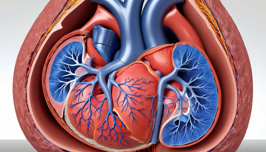
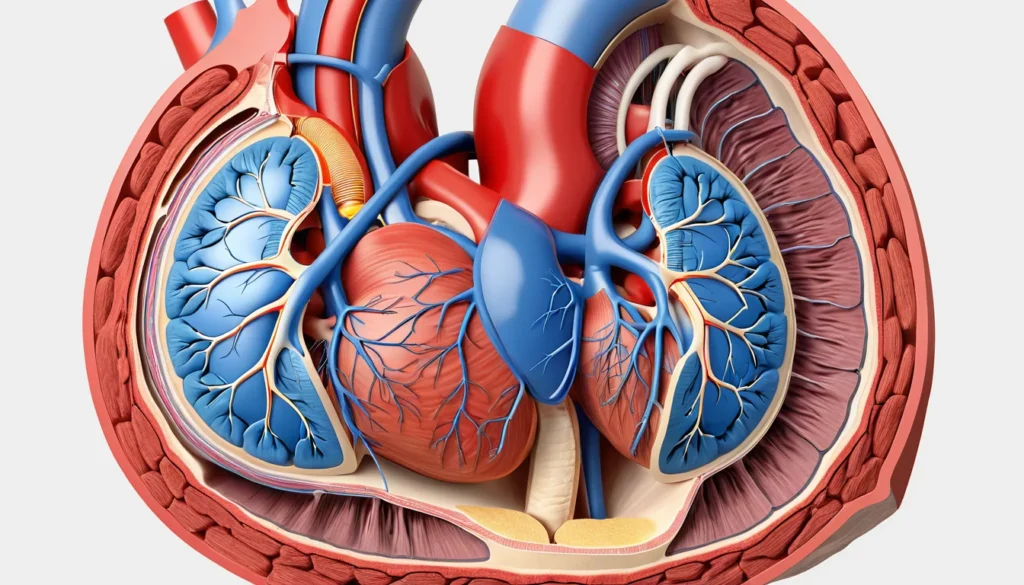
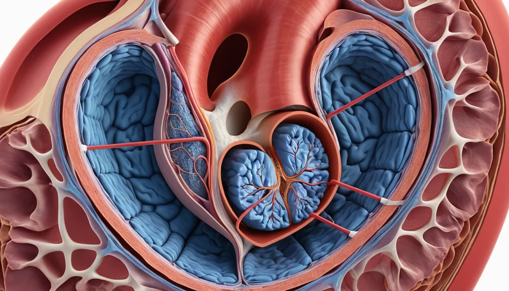
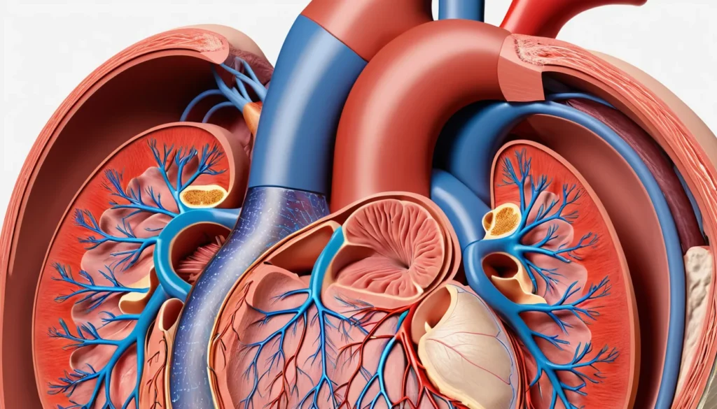
Your heart muscle normally stretches and relaxes with every heartbeat. In restrictive cardiomyopathy, the muscle loses this flexibility and becomes rigid. The ventricles can still squeeze and pump, but they cannot fill properly because the walls are too stiff.
When the ventricles cannot fill with enough blood, pressure builds up. Blood backs up into the circulatory system, affecting organs throughout the body. Over time, most people with this condition develop heart failure — a state where the heart can no longer meet the body’s needs for oxygen and nutrients.
Restrictive cardiomyopathy is rare in Canada and around the world. It is different from two other common types of cardiomyopathy. Dilated cardiomyopathy involves a weakened and stretched heart muscle. Hypertrophic cardiomyopathy involves a thickened, enlarged heart muscle. You can learn more about heart muscle diseases from Mayo Clinic’s overview of cardiomyopathy.
What Causes Restrictive Cardiomyopathy?
In many cases, doctors cannot find a clear cause. When no cause is identified, it is called idiopathic restrictive cardiomyopathy. However, several known conditions can trigger it.
Protein and Mineral Deposits
Amyloidosis is one of the more common causes. It involves abnormal protein deposits building up in the heart muscle, making it stiff. Haemochromatosis causes excess iron to accumulate in the heart tissue, damaging the muscle over time. Both conditions interfere with the heart’s normal structure and function.
Inflammatory Conditions
Sarcoidosis is a chronic disease where small clusters of inflammatory cells — called granulomas — form in organs and tissues. When granulomas develop in the heart, they disrupt its structure and ability to function normally. The lungs are also commonly affected.
Cancer Treatments
Radiation therapy and chemotherapy used to treat certain cancers can damage the heart muscle. This damage may lead to restrictive cardiomyopathy years after treatment ends. Therefore, people who have undergone cancer treatment should have regular heart check-ups as part of their follow-up care.
Immune and Genetic Causes
Löffler syndrome (also called hypereosinophilic syndrome) and endomyocardial fibrosis are conditions where a type of white blood cell called eosinophils causes scarring of the heart tissue. In addition, certain inherited metabolic disorders — such as Gaucher disease and Fabry disease — can cause restrictive cardiomyopathy through genetic pathways. If you have a family history of heart muscle disease, inform your family doctor so they can arrange appropriate screening.
Recognising the Symptoms of Restrictive Cardiomyopathy
The symptoms of restrictive cardiomyopathy closely resemble those of heart failure. They often develop gradually and can be easy to overlook at first. Recognising them early gives you the best chance at effective treatment.
Common Symptoms
Swelling in the legs, ankles, and feet due to fluid retention
Abdominal swelling and discomfort caused by fluid build-up (ascites) or blood backing up into the liver
Shortness of breath during everyday activities like walking or climbing stairs
Dry, persistent cough that worsens when lying down — it may wake you at night with a feeling of breathlessness
Dizziness, fatigue, and fainting — lightheadedness or brief loss of consciousness can occur
Heart palpitations — an awareness of irregular or rapid heartbeats
Frequent nighttime urination, which happens as the body tries to clear excess fluid while at rest
Furthermore, people with idiopathic restrictive cardiomyopathy may experience clot-related complications — such as stroke — before other symptoms appear. This makes early detection especially important.
Signs of a Medical Emergency
Restrictive cardiomyopathy can lead to acute heart failure, caused by sudden fluid build-up in the lungs (pulmonary oedema). This is a medical emergency. Call 911 immediately if you or someone near you experiences:
Severe difficulty breathing or a feeling of suffocation
Rapid or irregular heartbeat
Coughing up frothy, pink-tinged mucus
Acute heart failure can be triggered by eating too much salt, drinking large amounts of fluid, missing medications, or a sudden heart rhythm problem. It is distinct from chronic heart failure, which develops slowly over time.
Serious Complications to Be Aware Of
Beyond heart failure, restrictive cardiomyopathy can lead to other life-threatening complications. Understanding these risks helps you and your healthcare team make informed decisions about monitoring and treatment.
Blood Clots and Embolism
Blood clots can form inside the heart and break free into the bloodstream. A travelling clot — called an embolus — can block blood vessels in critical organs. Depending on where it lodges, it can cause:
A stroke, if it reaches the brain
A heart attack, if it blocks a coronary artery
Pulmonary embolism, if it travels to the lungs
Tissue damage in other organs and limbs
Arrhythmias and Sudden Cardiac Death
Abnormal heart rhythms — called arrhythmias — are a common complication. Atrial fibrillation is particularly frequent. In some cases, dangerous arrhythmias can lead to sudden cardiac death. For this reason, people with restrictive cardiomyopathy are closely monitored by a cardiologist. Learn more about arrhythmias from Health Canada’s heart disease resources.
How Is Restrictive Cardiomyopathy Diagnosed?
Diagnosing restrictive cardiomyopathy involves a combination of your personal and family medical history, a physical examination, and several tests. Early diagnosis leads to better symptom management and improved quality of life.
Physical Examination
Your doctor will listen to your heart and lungs with a stethoscope. Unusual heartbeats, heart murmurs, or a “gallop” sound may point to heart wall or valve problems. Crackling sounds in the lungs may signal fluid build-up. Your doctor will also check for swelling in your legs and look for signs of fluid retention elsewhere in the body.
Some cardiologists recommend regular heart monitoring for older adults — especially those over 60 — who have heart failure symptoms but a relatively normal heart size. Research suggests restrictive cardiomyopathy may be more common in this age group than previously thought. However, this condition can affect people of any age.
Diagnostic Tests
Your doctor may order several tests to confirm the diagnosis and identify the underlying cause:
Electrocardiogram (ECG) — records the heart’s electrical activity and detects rhythm problems
Echocardiogram — uses ultrasound to show the heart’s structure and how well it fills and pumps
Chest X-ray — checks for an enlarged heart or fluid in the lungs
MRI of the heart — provides detailed images of the heart muscle and can detect scarring or deposits
Blood tests — look for signs of infection, inflammation, iron overload, or protein abnormalities
Cardiac biopsy — a small sample of heart tissue may be taken to look for amyloid protein, iron deposits, or granulomas
A correct diagnosis often requires input from both a family doctor and a specialist cardiologist. Provincial health plans in most Canadian provinces cover these diagnostic tests when referred by a physician. For more information on heart failure diagnosis, visit Healthline’s guide to restrictive cardiomyopathy.
Treatment Options for Restrictive Cardiomyopathy
There is no cure for most forms of restrictive cardiomyopathy. However, treatment can significantly ease symptoms and reduce the risk of complications. Your treatment plan will depend on the underlying cause and how far the disease has progressed.
Medications
Doctors commonly prescribe several types of medication to manage symptoms:
Diuretics (water pills) — help remove excess fluid from the body, reducing swelling and lung congestion
Blood thinners (anticoagulants) — reduce the risk of blood clots forming in the heart
Anti-arrhythmic drugs — help control irregular heart rhythms
Medications targeting the underlying cause — for example, treatment for amyloidosis, iron chelation therapy for haemochromatosis, or corticosteroids for sarcoidosis
Lifestyle Adjustments
Alongside medication, certain lifestyle changes help protect your heart. Reducing your daily salt intake can prevent fluid build-up. Monitoring your fluid intake and your weight daily can help detect early signs of worsening heart failure. Light physical activity, as guided by your healthcare team, may also improve overall heart health.
Advanced Treatments
In severe cases, a pacemaker or implantable defibrillator (ICD) may be needed to manage dangerous heart rhythms. For end-stage restrictive cardiomyopathy, a heart transplant may be considered, particularly in younger patients without other serious health conditions. Your cardiologist will discuss whether these options are appropriate for your situation.
When to See a Doctor
If you notice any symptoms of restrictive cardiomyopathy — such as unexplained swelling, shortness of breath, persistent fatigue, or irregular heartbeats — do not wait. Book an appointment with your family doctor as soon as possible.
If you do not have a family doctor, a walk-in clinic can assess your symptoms and refer you to a cardiologist if needed. Most provincial health plans cover specialist referrals when recommended by a primary care provider.
If symptoms come on suddenly and are severe — particularly difficulty breathing, chest pain, or fainting — call 911 or go to your nearest emergency department immediately. Acute heart failure is a medical emergency.
Always speak with a qualified healthcare professional before making any changes to your heart health routine or medication. This article is for informational purposes only and does not replace personalised medical advice.
Frequently Asked Questions About Restrictive Cardiomyopathy
What is the life expectancy for someone with restrictive cardiomyopathy?
Life expectancy with restrictive cardiomyopathy varies depending on the underlying cause and how early it is diagnosed. Idiopathic cases can progress more rapidly, while forms caused by treatable conditions — like haemochromatosis — may improve with targeted therapy. Your cardiologist can give you a clearer picture based on your individual situation.
Is restrictive cardiomyopathy hereditary?
In some cases, restrictive cardiomyopathy is linked to inherited metabolic disorders such as Fabry disease or Gaucher disease. If a close family member has been diagnosed with a heart muscle disease, inform your family doctor so appropriate genetic counselling or screening can be arranged. Not all cases have a genetic cause.
How is restrictive cardiomyopathy different from other types of cardiomyopathy?
Restrictive cardiomyopathy is characterised by a stiff heart muscle that cannot relax and fill properly, while dilated cardiomyopathy involves a weakened, enlarged heart, and hypertrophic cardiomyopathy involves an abnormally thickened muscle. Each type affects the heart differently and requires a distinct approach to treatment. An echocardiogram is usually the key test used to tell them apart.
Can restrictive cardiomyopathy be cured?
Currently, there is no cure for most forms of restrictive cardiomyopathy. However, treatment can effectively manage symptoms and slow the progression of the disease. In cases caused by a specific treatable condition — such as amyloidosis or iron overload — targeting the underlying cause can significantly improve heart function.
What are the early warning signs of restrictive cardiomyopathy?
Early signs of restrictive cardiomyopathy include unexplained tiredness, swelling in the legs or ankles, shortness of breath during mild activity, and a dry cough that worsens when lying down. Because these symptoms are easy to dismiss, many people