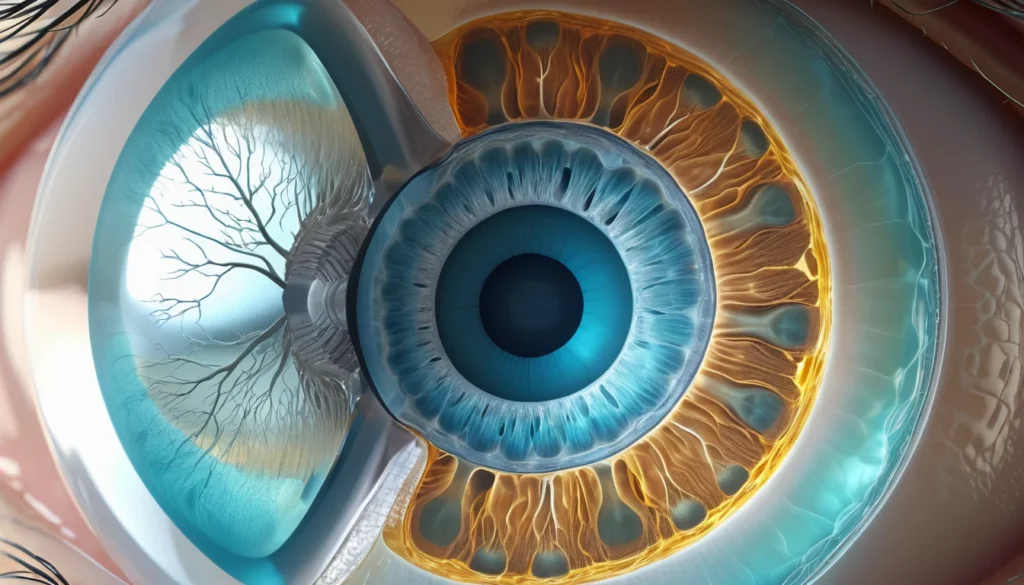
Keratoconus is an eye condition where the cornea — the clear, dome-shaped front surface of the eye — gradually thins and bulges outward into a cone shape. This shape change distorts vision and can make everyday tasks like reading or driving increasingly difficult. In Canada, many people live with this condition for years before receiving a proper diagnosis. This article explains what keratoconus is, what causes it, how it is treated, and when you should speak with a healthcare provider.
What Is Keratoconus?
The cornea normally holds a rounded shape. It focuses light onto the retina at the back of the eye, producing clear vision. In keratoconus, the structural proteins — called collagen — inside the cornea weaken over time.
As the collagen weakens, the cornea can no longer hold its round shape. It begins to thin and bulge forward, forming a cone-like curve. This irregular shape scatters light instead of focusing it, causing blurry and distorted vision.
Keratoconus typically begins in the teen years or early twenties. It usually affects both eyes, though often one eye is more severely affected than the other. The condition tends to progress slowly over many years before stabilising in most people.
What Causes Keratoconus?
The exact cause of keratoconus is not fully understood. However, researchers believe a combination of genetic and environmental factors plays a role. According to Mayo Clinic’s overview of keratoconus, abnormalities in corneal collagen are central to how the condition develops.
There is a strong hereditary component. If a parent or sibling has keratoconus, your risk of developing it is higher. For this reason, family members of someone diagnosed with the condition are encouraged to have their eyes examined regularly.
Associated Risk Factors
Several factors may increase the likelihood of developing keratoconus. These include:
Genetics: A family history of keratoconus significantly raises your risk.
Down syndrome (Trisomy 21): People with Down syndrome have a higher rate of keratoconus.
Atopy: This is a tendency toward allergic conditions such as asthma, eczema, or hay fever. Frequent eye rubbing, common in people with allergies, may accelerate corneal thinning.
Eye rubbing: Vigorous and repeated rubbing of the eyes over time may damage the corneal tissue.
Connective tissue disorders: Conditions that affect the body’s structural proteins may also be linked.
It is important to note that having one or more of these risk factors does not guarantee you will develop keratoconus. However, being aware of them allows you and your eye doctor to monitor your eye health more closely.
Recognising the Symptoms of Keratoconus
In its early stages, keratoconus can be easy to miss. The changes in vision happen gradually, and many people assume they simply need a stronger glasses prescription. Over time, however, the symptoms become more noticeable and harder to correct with standard eyewear.
Early Symptoms
The first sign of keratoconus is usually a form of astigmatism that appears later in life than typical astigmatism. Astigmatism means the eye does not focus light evenly, causing blurry or distorted vision. In keratoconus, this astigmatism is irregular, meaning it does not follow a predictable pattern that standard glasses can fully correct.
Other early symptoms include:
Slightly blurred or distorted vision
Increased sensitivity to light and glare
Frequent changes in your glasses or contact lens prescription
Difficulty seeing clearly at night
Advanced Symptoms
As keratoconus progresses, the cornea continues to thin and bulge further. The astigmatism becomes more severe and irregular. Standard glasses may no longer provide adequate vision correction at this stage.
In more advanced cases, clouding or scarring can develop in and around the distorted area of the cornea. This scarring further reduces visual clarity. Some people experience sudden episodes of worsening vision caused by a condition called acute hydrops, where fluid enters the cornea through small cracks.
For a broader understanding of how corneal conditions affect vision, Healthline’s guide to keratoconus offers a helpful and accessible overview.
How Is Keratoconus Diagnosed?
An eye specialist — called an optometrist or ophthalmologist — can diagnose keratoconus through a detailed eye examination. A standard vision test alone is not enough. Specific tests are needed to measure the shape and thickness of the cornea.
Common Diagnostic Tests
The most important test is corneal topography. This painless scan creates a detailed map of the cornea’s surface. It can detect even subtle irregularities in shape that would not be visible during a routine exam.
Additional tests may include:
Pachymetry: This measures the thickness of the cornea at various points.
Slit-lamp examination: A special microscope allows the doctor to examine the layers of the cornea closely.
Refraction testing: This checks how the eye bends light, helping to identify irregular astigmatism.
Early diagnosis is very important. Catching keratoconus before significant thinning occurs gives doctors more options for managing and slowing its progression.
Treatment Options for Keratoconus
The good news is that keratoconus is a treatable condition. The right treatment depends on how advanced the condition is and how quickly it is progressing. Treatment generally moves through several stages as the condition develops.
Glasses and Standard Contact Lenses
In the early stages, regular prescription glasses can correct the mild astigmatism caused by keratoconus. As the condition progresses and the astigmatism becomes more irregular, glasses may no longer provide sharp vision.
At this point, specially designed contact lenses become the preferred option. Rigid gas-permeable lenses, or scleral lenses, vault over the irregular corneal surface. They create a smooth optical surface and significantly improve vision. Many Canadians with moderate keratoconus manage their condition successfully with these lenses.
Corneal Cross-Linking
Corneal cross-linking is a procedure designed to slow or stop the progression of keratoconus. A doctor applies riboflavin (vitamin B2) eye drops to the cornea and then uses ultraviolet light to strengthen the collagen bonds within the corneal tissue.
This procedure does not reverse existing damage, but it can halt further thinning and bulging. It is most effective when keratoconus is caught and treated early. Cross-linking is available at specialised eye centres across Canada.
Corneal Transplant Surgery
In advanced cases where the cornea has thinned severely or developed significant scarring, a corneal transplant may be necessary. During this procedure, a surgeon replaces the damaged cornea with healthy donor corneal tissue.
Corneal transplants for keratoconus have very good outcomes. The vast majority of patients experience a meaningful improvement in vision following the procedure. However, recovery can take months, and patients often still need glasses or contact lenses afterwards to fine-tune their vision.
Canada has a well-established tissue donation and transplant network. Your ophthalmologist can refer you to an appropriate surgical centre through your provincial health system.
Living With Keratoconus in Canada
Managing keratoconus is an ongoing process, but many Canadians live full, active lives with this condition. Regular eye check-ups are essential for monitoring any changes in the cornea. Your optometrist or ophthalmologist will advise you on how often you should be seen.
Avoiding eye rubbing is one of the most important lifestyle adjustments. If you have allergies that cause itchy eyes, speak with your family doctor about managing them effectively. Treating the underlying allergy can reduce the urge to rub and may help protect your corneal tissue.
Most provincial health plans in Canada cover visits to optometrists and ophthalmologists, though coverage varies by province. Contact your provincial health authority or speak with your family doctor to understand what is covered under your plan. Some treatments, such as specialised contact lenses or cross-linking, may involve out-of-pocket costs that could be partially covered by extended health benefits.
For general guidance on eye health in Canada, the Health Canada website provides reliable, up-to-date information on accessing healthcare services across the country.
When to See a Doctor
You should see a healthcare provider if you notice any changes in your vision that cannot be corrected fully with your current glasses or contact lenses. Frequent prescription changes, blurry vision, or new sensitivity to light and glare are all reasons to seek an eye examination promptly.
Start by booking an appointment with your family doctor or visiting a walk-in clinic. They can refer you to an optometrist or ophthalmologist for a more detailed eye assessment. If you already see an eye specialist, contact their office directly.
If you have a family history of keratoconus, Down syndrome, or significant allergic conditions, mention this to your healthcare provider. This information helps them decide how closely to monitor your eye health over time. Early detection makes a significant difference in the range of treatment options available to you.
As always, consult your doctor or a qualified eye specialist before making any decisions about your eye health. The information in this article is for general educational purposes and is not a substitute for professional medical advice.
Frequently Asked Questions About Keratoconus
Can keratoconus lead to blindness?
Keratoconus does not typically cause total blindness, but it can lead to severe vision impairment if left untreated. With proper management — including contact lenses, cross-linking, or a corneal transplant — most people with keratoconus maintain functional vision throughout their lives. Early diagnosis and treatment are key to preventing serious vision loss.
Is keratoconus hereditary?
Yes, keratoconus has a strong hereditary component, meaning it can run in families. If a parent or sibling has keratoconus, you have a higher chance of developing it yourself. Regular eye exams are recommended for family members of anyone diagnosed with this condition.
At what age does keratoconus typically start?
Keratoconus most often begins during the teenage years or early twenties. The condition tends to progress slowly over the next one to two decades before stabilising in most people around the age of 40. However, onset and progression vary from person to person.
Can rubbing your eyes make keratoconus worse?
Yes, frequent and vigorous eye rubbing is believed to accelerate the progression of keratoconus by further weakening the corneal tissue. People who have keratoconus or are at risk for it should avoid rubbing their eyes as much as possible. If allergies are causing eye irritation, speak with your doctor about effective treatment options.
Is corneal cross-linking covered by provincial health plans in Canada?
Coverage for corneal cross-linking varies by province and individual health plan. Some provincial health programmes cover the procedure when it is deemed medically necessary, while others may require partial or full out-of-pocket payment. Contact your provincial health authority or your family doctor to find out what is available under your specific plan.
How successful is corneal transplant surgery for keratoconus?
Corneal transplant surgery for keratoconus has a very high success rate, with the majority of patients experiencing significant improvement in vision. Most people still need glasses or specially fitted contact lenses after surgery to achieve their best possible vision. Recovery can take several months, so follow-up care with your eye specialist is essential.
Key Takeaways
Keratoconus is a progressive eye condition where the cornea thins and bulges into a cone shape, distorting vision.
The exact cause is unknown, but genetics, atopy, and eye rubbing are known risk factors.
Symptoms include irregular astigmatism, blurry vision, light sensitivity, and frequent prescription changes.
Treatment ranges from glasses and specialised contact lenses in early stages to corneal cross-linking or transplant surgery in advanced cases.
Early diagnosis is essential — it expands your treatment options and can slow the progression of the condition significantly.
Canadians should speak with their family doctor, optometrist, or ophthalmologist if they notice changes in their vision.
Always consult a qualified healthcare professional for advice specific to your personal health situation.