A chest ultrasound is a safe, fast, and radiation-free way for doctors to look at your lungs and the structures around them. Over the past decade, it has become one of the most useful tools in respiratory medicine. In this article, we explain what a chest ultrasound is, when your doctor might recommend one, and what to expect during the procedure.
What Is a Chest Ultrasound?
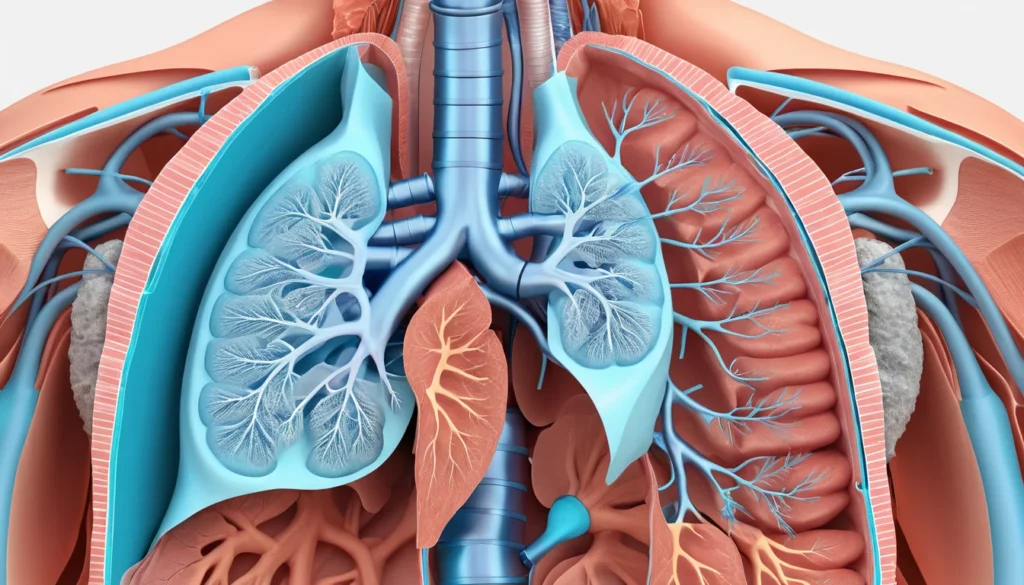
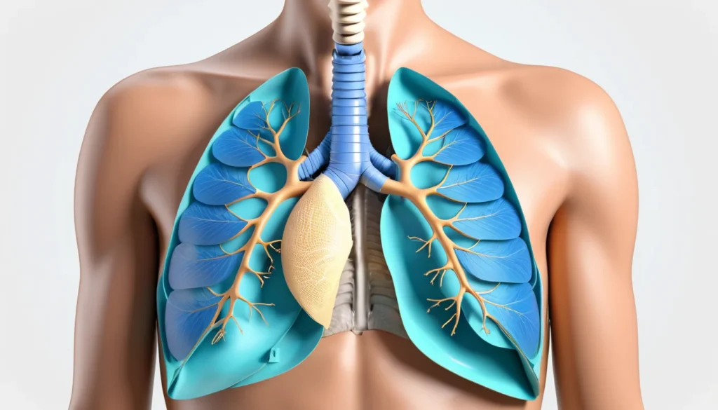
A chest ultrasound uses sound waves to create real-time images of the structures inside your chest. These include your lungs, the lining around them (called the pleura), your diaphragm, and nearby blood vessels.
Unlike X-rays or CT scans, a chest ultrasound does not use radiation. A small handheld device called a transducer is gently pressed against your skin. It sends sound waves into your body and picks up the echoes that bounce back, building a picture of what is inside.
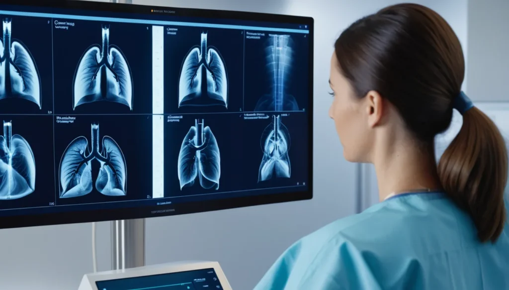
For many years, chest X-rays and CT scans were the main tools doctors used to check the lungs. However, chest ultrasound has grown in popularity because it is portable, affordable, and can be done right at a patient’s bedside. According to the World Health Organization, lung diseases are among the leading causes of illness worldwide — making better diagnostic tools more important than ever.
What Can a Chest Ultrasound See?
A chest ultrasound is especially useful for looking at the pleural space — the thin gap between your lungs and the chest wall. It can spot fluid, air, or abnormal tissue in that area.
Because air inside the lungs blocks sound waves, a chest ultrasound cannot image deep lung tissue as clearly as a CT scan can. However, it is highly effective for evaluating the outer surface of the lungs and surrounding structures.
Conditions a Chest Ultrasound Can Help Diagnose
Your doctor may use a chest ultrasound to investigate or monitor a wide range of conditions, including:
Pleural effusion — a build-up of fluid around the lungs
Pneumothorax — a collapsed lung caused by air in the pleural space
Pneumonia — infection and inflammation in the lungs
Lung abscesses — pockets of infection in the lung tissue
Lung tumours — both benign and malignant masses near the lung surface
Rib fractures — breaks in the bones of the chest wall
Diaphragmatic hernia — when organs push through the diaphragm
Pulmonary embolism — a blood clot in the lungs
Heart failure — which can cause fluid to build up around the lungs
Arteriovenous malformations — abnormal connections between arteries and veins, often near the outer lung
In addition, chest ultrasound can help distinguish between fluid collections near the lungs versus those below the diaphragm. This detail matters a great deal when planning treatment.
When Is a Chest Ultrasound Recommended?
A chest ultrasound is often recommended when a doctor needs a quick and reliable answer — and waiting for a CT scan is not practical. It is particularly valuable in emergency or bedside settings, such as in hospital intensive care units or emergency departments.
Your doctor might recommend a chest ultrasound in these situations:
You have unexplained shortness of breath or chest pain
A chest X-ray shows something unclear that needs further investigation
A CT scan or X-ray is not available or not safe for you right now
Your doctor needs to guide a procedure, such as draining fluid from around the lungs (called thoracentesis)
You are being monitored for a known lung or pleural condition
There is a concern about a lung tumour spreading into the chest wall
Furthermore, chest ultrasound is used to guide interventional procedures safely. For example, when a doctor needs to insert a chest drain or perform a pleural biopsy, ultrasound helps them place instruments accurately and reduces the risk of complications like pneumothorax.
Advantages of Chest Ultrasound
There are many reasons why chest ultrasound has become a go-to tool for respiratory specialists and emergency doctors alike. Here are the key benefits:
No radiation — safe for pregnant women, children, and patients who need repeated imaging
Portable — the equipment can be brought directly to a patient’s bedside, even in the ICU
Real-time imaging — doctors see results immediately, with no waiting for lab processing
Non-invasive — no needles, no injections, no preparation required
Cost-effective — generally less expensive than CT scanning
More precise for fluid location — better than a physical exam alone for pinpointing where fluid sits
Lower risk during procedures — reduces the chance of pneumothorax when draining pleural fluid
Quick to learn — medical professionals can be trained in basic chest ultrasound relatively quickly
As a result, many Canadian hospitals now use chest ultrasound as a first-line tool in emergency and critical care settings. Healthline explains more about how chest ultrasound works and what patients can expect.
Limitations of Chest Ultrasound
Like any medical test, a chest ultrasound has its limitations. It is important to understand what it cannot do as well as what it can.
Limited view of deep lung tissue — air inside the lungs blocks sound waves, so a CT scan is still better for imaging the full lung interior
Artefacts from air — air interference can create misleading images that require expert interpretation
Ribs block the view — the rib cage can hide some abnormalities from the ultrasound beam
Less useful for complex interventions — for complicated surgical procedures, CT guidance may still be preferred
Therefore, a chest ultrasound is often used alongside other imaging tests rather than replacing them entirely. Your doctor will choose the right combination of tests based on your specific situation.
What to Expect During the Procedure
A chest ultrasound is a straightforward and painless procedure. There is no special preparation needed beforehand — you do not need to fast or stop any medications.
How You Will Be Positioned
Your positioning depends on what your doctor is looking for. In most cases, you will sit upright in a chair with your arms resting on a table or chair back. This position allows fluid to settle naturally due to gravity, making it easier to see.
If you are unable to sit up — for example, if you are a hospital patient on bed rest — the exam can be done while you lie down with the head of the bed slightly raised. The sonographer will place your arm across your chest to improve access to the side being examined.
How the Scan Is Performed
The sonographer or doctor will apply a clear gel to your skin. This gel helps the transducer make proper contact and transmit sound waves effectively.
The transducer is then moved across your chest in different directions. First, it is held lengthwise for a general overview of the chest. Next, the doctor may tilt it sideways to focus on a specific area of concern. The whole exam usually takes between 15 and 30 minutes.
The machine uses several different modes to capture information. Brightness mode (B-mode) creates the familiar two-dimensional images. Motion mode (M-mode) tracks movement of structures over time. Doppler mode detects blood flow, which helps doctors tell the difference between a fluid collection and a vascularised mass.
Before the exam begins, you will be asked to sign an informed consent form. This is standard practice in Canadian healthcare. Your doctor or the technician will explain the purpose of the test, the technique, and any potential risks — which are minimal for this type of imaging.
When to See a Doctor
You should speak with your family doctor or visit a walk-in clinic if you experience any of the following symptoms:
Unexplained shortness of breath, especially at rest
Persistent cough lasting more than three weeks
Chest pain or tightness
Coughing up blood
Unexplained fatigue combined with breathing difficulty
A recent respiratory illness that is not improving
Your family doctor can refer you for a chest ultrasound through your provincial health plan if they feel it is needed. In urgent cases, an emergency department can perform the scan right away. Health Canada outlines how to access diagnostic services in your province.
It is always best to discuss any breathing or chest concerns with a qualified healthcare provider. This article is for general information only and is not a substitute for professional medical advice.
Frequently Asked Questions About Chest Ultrasound
Is a chest ultrasound the same as a chest X-ray?
No, a chest ultrasound and a chest X-ray are different tests. A chest X-ray uses radiation to create a single flat image, while a chest ultrasound uses sound waves to produce real-time moving images. A chest ultrasound is particularly better at detecting fluid around the lungs and guiding certain procedures.
Is a chest ultrasound safe?
Yes, a chest ultrasound is considered very safe. It does not use radiation, making it suitable for pregnant women, children, and patients who need frequent monitoring. There are no known risks associated with diagnostic ultrasound used at standard medical settings.
How long does a chest ultrasound take?
A chest ultrasound typically takes between 15 and 30 minutes, depending on what your doctor is looking for. No preparation is required beforehand, so you can return to normal activities immediately after. Results are often available right away since the images are seen in real time.
Can a chest ultrasound detect lung cancer?
A chest ultrasound can help detect tumours that are located near the surface of the lung or in the chest wall. It can also help distinguish between benign and malignant masses using Doppler blood flow analysis. However, a CT scan is generally more reliable for a full evaluation of lung cancer, and your doctor may order both tests.
Does provincial health insurance in Canada cover chest ultrasound?
In most provinces, a chest ultrasound ordered by a physician is covered under your provincial health plan when it is medically necessary. Coverage details vary by province, so it is best to confirm with your family doctor or provincial health authority. In most cases, you will not pay out of pocket for a referred diagnostic ultrasound.
What is the difference between a chest ultrasound and a CT scan of the chest?
A chest ultrasound uses harmless sound waves and is best for imaging the pleura, chest wall, and fluid collections in real time. A CT scan uses radiation and provides a much more detailed view of the entire lung tissue and internal structures. Doctors often use a chest ultrasound first because it is faster and radiation-free, then follow up with a CT scan if more detail is needed.
Key Takeaways
A chest ultrasound uses sound waves to image the lungs, pleura, diaphragm, and chest wall in real time.
It is safe, radiation-free, portable, and cost-effective — making it ideal for bedside use in Canadian hospitals.
It is especially useful for detecting pleural fluid, pneumothorax, pneumonia, and guiding drainage procedures.
It has limitations — it cannot image deep lung tissue as well as a CT scan, and air inside the lungs can interfere with the signal.
No preparation is needed, and the procedure is painless and quick.
If you have unexplained breathing problems or chest symptoms, speak with your family doctor or visit a walk-in clinic. They can refer you for imaging through your provincial health plan.
This article is for informational purposes only. Always consult a qualified healthcare provider for diagnosis and treatment.