Brain sand, also known by its medical name acervulus cerebri, refers to tiny calcium-containing granules that build up in certain parts of the brain over time. These deposits are most commonly found in the pineal gland, a small pea-sized gland located deep inside the brain. For most people, brain sand is completely harmless and causes no symptoms at all. In this article, we explain what brain sand is, where it forms, why it happens, and what — if anything — you need to do about it.
What Is Brain Sand?
Brain sand is the common name for small, gritty calcium deposits that form inside brain tissue. The medical term, acervulus cerebri, comes from the Latin word for “little heap.” These deposits look like grains of sand under a microscope, which is how they got their name.
These calcium granules are not a disease. They are a normal finding in many adults, and they become more common as people age. Researchers have found brain sand in humans of all backgrounds, and it shows up regularly on brain scans performed for other reasons.
In most cases, a doctor will notice brain sand on an X-ray, CT scan, or MRI and simply note it as a benign — meaning harmless — incidental finding. No treatment is needed unless another underlying condition is present.
Where Does Brain Sand Form in the Brain?
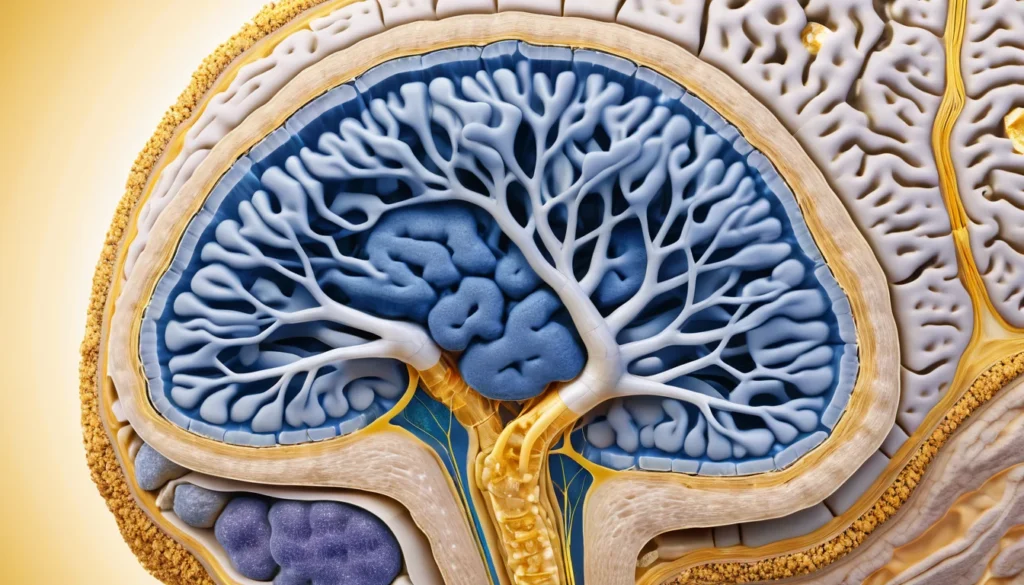
Brain sand most often forms in the pineal gland. The pineal gland sits near the centre of the brain and plays a role in regulating sleep by producing a hormone called melatonin. It is one of the most common sites for calcium deposits in the brain.
However, brain sand can also appear in other brain structures. These include the choroid plexus, which produces the fluid that cushions the brain, and the habenula, a small region involved in processing emotions and regulating sleep-wake cycles.
In addition, calcium deposits may occasionally appear in the dura mater, the tough outer membrane that covers the brain and spinal cord. All of these locations are considered normal sites for minor calcification as the brain ages.
Brain Sand vs. Abnormal Brain Calcification
It is important to understand the difference between normal brain sand and abnormal brain calcification. Normal brain sand is small, contained, and not linked to any disease process. Abnormal calcification, on the other hand, may be larger, appear in unusual locations, or be associated with conditions such as infections, metabolic disorders, or tumours.
Your doctor is the best person to determine whether a calcium deposit seen on a brain scan is typical brain sand or something that needs further investigation. In most cases, it is completely normal.
Why Does Brain Sand Form?
The exact reason brain sand forms is not fully understood. However, researchers believe it is a natural part of ageing. As the brain gets older, certain cells begin to deposit calcium as part of their normal life cycle.
The pineal gland, in particular, tends to calcify gradually over a person’s lifetime. Studies suggest that pineal calcification can begin as early as the teenage years, and it becomes increasingly common after age 40. By age 60, a majority of adults show some degree of pineal calcification on brain imaging.
Furthermore, some researchers believe that brain sand may be linked to the body’s regulation of calcium and phosphate levels. Changes in these mineral levels over time may contribute to the gradual build-up of calcium in brain tissue. However, this does not mean that having brain sand indicates a problem with your bones or your calcium levels.
Factors That May Influence Brain Sand Formation
While ageing is the primary factor, a few other influences may play a role. These include:
Age: Brain sand becomes more common and more visible with age.
Genetics: Some families may show a higher tendency toward brain calcification.
Hormonal changes: The pineal gland is a hormonal organ, and shifts in hormone levels over a lifetime may affect calcification rates.
Geographic and dietary factors: Some studies suggest that mineral intake, including calcium and fluoride, may play a small role.
It is worth noting that none of these factors are under your direct control in a meaningful way. Brain sand is not caused by a poor diet or an unhealthy lifestyle. It is simply a natural process that happens in many people.
Does Brain Sand Cause Any Symptoms?
In the vast majority of cases, brain sand causes no symptoms whatsoever. Most people who have it never know it is there. It is typically discovered by accident during a brain scan ordered for a completely different reason, such as a headache investigation or a head injury assessment.
Because the deposits are tiny and do not usually interfere with brain function, they do not cause headaches, memory problems, vision changes, or any other neurological symptoms on their own.
However, if the pineal gland becomes very heavily calcified, some researchers have questioned whether it could affect melatonin production and, therefore, sleep quality. The evidence on this is not conclusive, and most sleep experts point to other causes — such as stress, screen time, or irregular schedules — as far more significant contributors to poor sleep.
When Brain Sand Might Be Relevant
In rare cases, large or unusual calcium deposits in the brain can be a sign of an underlying medical condition. For example, certain infections, such as toxoplasmosis or tuberculosis, can leave calcium deposits in the brain after they heal. Conditions like hypoparathyroidism — a disorder affecting the parathyroid glands — can also cause abnormal brain calcification.
As a result, if your doctor sees a calcium deposit on a brain scan, they may ask you a few questions or order additional tests to rule out these less common causes. This is simply good medical practice, not a cause for alarm.
How Is Brain Sand Detected?
Brain sand is detected through medical imaging. The most common ways it shows up include:
CT scan (computed tomography): This is the most sensitive imaging tool for detecting calcium deposits. Even very small granules of brain sand are visible on a CT scan.
X-ray: Larger calcium deposits in the pineal gland may appear on a plain skull X-ray.
MRI (magnetic resonance imaging): MRI is excellent for showing brain tissue in detail, though it is less sensitive than CT for detecting small calcium deposits.
In Canada, these imaging tests are typically ordered by your family doctor or a specialist when there is a medical reason to investigate. Provincial health plans generally cover brain imaging when it is medically necessary. Your doctor will refer you through the appropriate channels based on your symptoms and history.
You can learn more about how brain imaging works by visiting the Mayo Clinic’s guide to CT scans.
Brain Sand and the Pineal Gland: What You Should Know
The pineal gland has fascinated scientists and philosophers for centuries. The 17th-century philosopher René Descartes famously called it the “seat of the soul.” Today, we know it is a small endocrine gland that produces melatonin and helps regulate the body’s internal clock, known as the circadian rhythm.
Pineal calcification — or brain sand in the pineal gland — is so common that radiologists use it as a landmark on brain scans. Because it sits at the exact centre of the brain, a calcified pineal gland helps doctors check whether the brain’s midline structures are properly aligned. A shift in the location of the calcified pineal gland can sometimes indicate swelling or a mass on one side of the brain.
Therefore, while brain sand itself is harmless, its presence on a scan can actually be useful to doctors as a reference point. For more detailed information about the pineal gland and its functions, you can visit Healthline’s overview of the pineal gland.
Does a Calcified Pineal Gland Affect Melatonin or Sleep?
This is a question many Canadians ask after learning they have pineal calcification. The short answer is: probably not significantly. While some studies have found a mild association between heavy pineal calcification and lower melatonin levels, the overall evidence is mixed and inconclusive.
Most people with brain sand sleep normally and have no issues with their circadian rhythm. If you are experiencing poor sleep, it is far more likely related to lifestyle factors, stress, or other health conditions than to brain sand. Talk to your family doctor if sleep problems are affecting your daily life.
When to See a Doctor
If you have been told you have brain sand or a calcified pineal gland after a brain scan, there is usually no reason to panic. In most cases, your doctor will simply monitor it or take note of it in your medical record.
However, you should speak with your family doctor or visit a walk-in clinic if you experience any of the following:
Severe or persistent headaches that are new or unusual for you
Vision changes, such as double vision or blurred sight
Significant changes in memory or thinking ability
Unexplained personality or behaviour changes
Seizures or episodes of confusion
Weakness or numbness on one side of the body
These symptoms are not caused by typical brain sand, but they may point to other neurological conditions that need prompt attention. Your family doctor can assess your symptoms and refer you to a neurologist or order imaging if needed. In Canada, most provincial health plans cover neurology referrals when there is a medical indication.
For general information on brain health and when to seek care, you can visit Health Canada’s official health information resources.
As always, please consult a qualified healthcare professional before drawing any conclusions about your own health based on information you read online. Every person’s situation is different, and only your doctor can give you advice tailored to your specific circumstances.
Frequently Asked Questions About Brain Sand
What is brain sand and is it dangerous?
Brain sand refers to tiny calcium deposits that form naturally in the brain, most often in the pineal gland. For the vast majority of people, brain sand is completely harmless and causes no symptoms. It is considered a normal part of ageing and is not dangerous on its own.
What causes brain sand to form?
Brain sand forms as a natural result of ageing, as certain brain cells gradually deposit calcium over time. The pineal gland is the most common site for this type of calcification. Factors such as genetics and mineral metabolism may also play a role, though the exact cause is not fully understood.
Does brain sand affect memory or brain function?
In the vast majority of cases, brain sand does not affect memory, thinking, or any other aspect of brain function. The deposits are very small and do not interfere with normal brain activity. If you are experiencing memory problems, it is important to speak with your family doctor to find the actual cause.
Can brain sand affect sleep or melatonin levels?
Some research has looked at whether pineal calcification, a form of brain sand, might reduce melatonin production and disrupt sleep. The evidence so far is mixed and not conclusive. Most sleep specialists attribute sleep problems to lifestyle, stress, and other health conditions rather than brain sand.
How is brain sand detected?
Brain sand is most often detected incidentally during a CT scan or MRI ordered for another reason, such as investigating headaches. CT scans are particularly sensitive at identifying calcium deposits in the brain. In Canada, your family doctor or specialist will order imaging if there is a medical reason to do so, and provincial health plans typically cover these tests.
Does brain sand require treatment?
In most cases, brain sand requires no treatment at all. Because it is a benign and natural finding, doctors typically just note it on your medical record. However, if calcium deposits in the brain are unusually large or appear in unexpected locations, your doctor may investigate further to rule out underlying conditions.
Key Takeaways
Brain sand (acervulus cerebri) is a collection of tiny calcium granules that form naturally in the brain, most often in the pineal gland.
It is a completely normal and common finding, especially in adults over 40.
Brain sand causes no symptoms in the vast majority of people and requires no treatment.
It is usually discovered by accident during a brain scan performed for another reason.
The pineal gland, where brain sand most commonly forms, regulates sleep through melatonin production — but brain sand does not significantly affect this function for most people.
If you have unusual neurological symptoms such as severe headaches, vision changes, or memory problems, see your family doctor or visit a walk-in clinic promptly.
Always consult a qualified healthcare professional for advice specific to your own health situation.