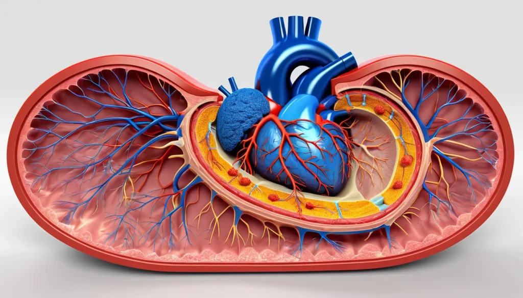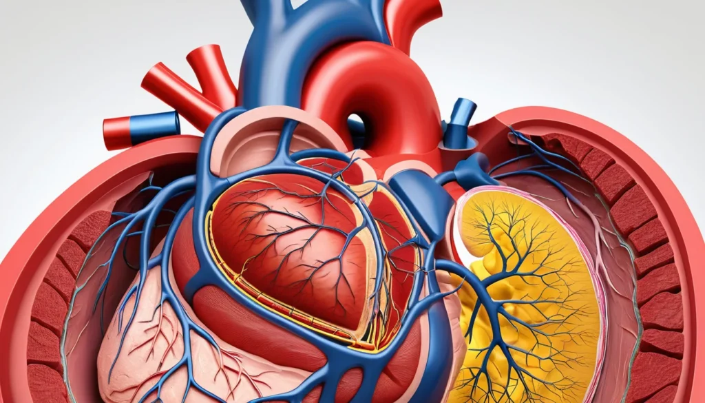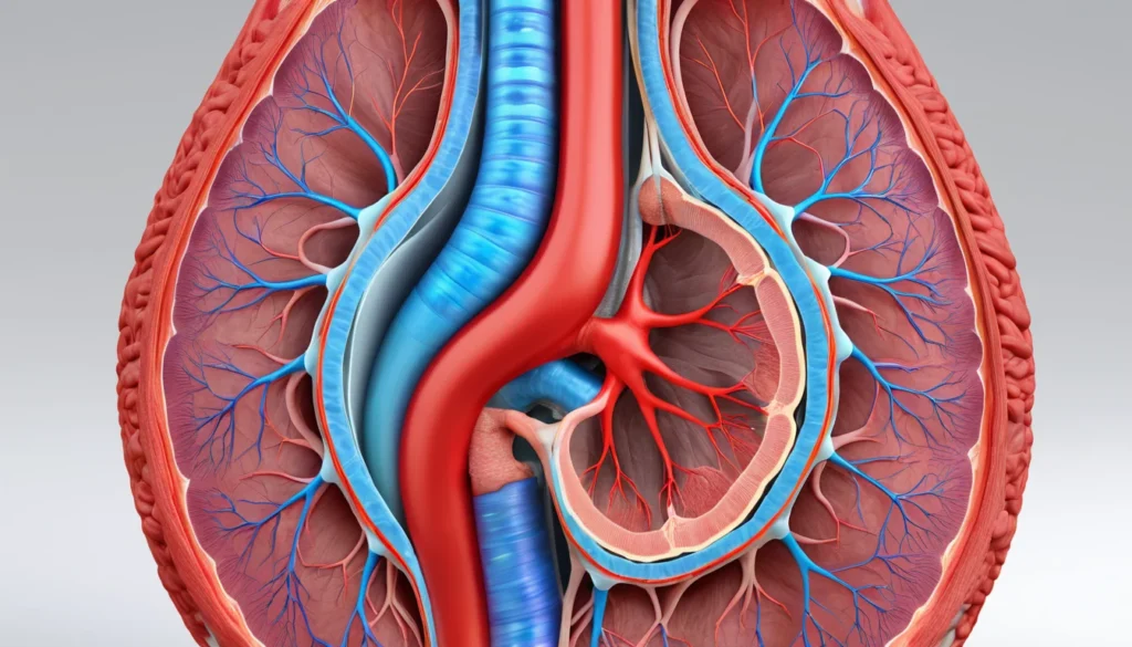
Aortic coarctation is a congenital heart defect where the aorta — the body’s main artery — becomes abnormally narrowed. This narrowing forces the heart to work much harder to pump blood through the body. It can cause high blood pressure in the upper body and reduced blood flow to the lower body. In this article, we explain what aortic coarctation is, what causes it, how it is diagnosed, and what treatment options are available to Canadians.
What Is Aortic Coarctation?
The aorta is the large artery that carries blood from your heart to the rest of your body. In aortic coarctation, a section of this artery is too narrow. This narrowing most often occurs just past the left subclavian artery, near a structure called the ductus arteriosus.
As a result, blood pressure rises in the upper body — including the arms and head — while the lower body receives less blood than it needs. This is called hypoperfusion. Think of it like a kinked garden hose: pressure builds up behind the kink, and water barely trickles out past it.
Most cases of aortic coarctation are present at birth. However, in rare cases, it can develop later in life due to aortic dissection caused by trauma. Learn more about aortic coarctation from the Mayo Clinic.
What Causes Aortic Coarctation?
Aortic coarctation develops during fetal growth, usually due to a problem with how the fifth and sixth aortic arches form. Experts have proposed two main theories to explain this.
The Ductal Tissue Theory
This theory suggests that smooth muscle from the ductus arteriosus migrates into the nearby aortic wall. When this tissue contracts after birth, it causes the aorta to narrow. This is currently the most widely accepted explanation.
The Haemodynamic Theory
This theory proposes that reduced blood flow during fetal development leads to underdevelopment of the aortic isthmus — the section most commonly affected. In other words, the aorta does not grow properly because it does not receive enough blood flow while the baby is developing.
In some cases, the elastic tissue in the aortic wall is also affected, especially when the ductus arteriosus and ligamentum arteriosum are located further away from the narrowing.
Risk Factors for Aortic Coarctation
Aortic coarctation accounts for roughly 5% of all congenital heart defects. It also causes approximately 7% of critical heart conditions in newborns. Understanding who is at higher risk can help with early detection.
Sex: Aortic coarctation is about twice as common in males as in females. However, among newborns, it affects both sexes equally.
Family history: Children whose parents have congenital heart defects face a higher risk of being born with similar conditions.
Turner syndrome: Girls born with Turner syndrome — a chromosomal condition — have a significantly higher risk of aortic coarctation.
Ethnicity: The condition appears more often in people of Caucasian background compared to those of Asian or African descent.
Other heart defects: Aortic coarctation frequently occurs alongside other heart problems, such as a bicuspid aortic valve or ventricular septal defect.
The age at which symptoms appear depends on how severe the narrowing is and whether other heart abnormalities are present at the same time.
Symptoms of Aortic Coarctation
Symptoms vary widely depending on the severity of the narrowing and the age of the person. Some people have no symptoms at all for many years. Others experience serious problems shortly after birth.
Symptoms in Newborns
In severe cases, symptoms appear within the first three weeks of life. These can include difficulty feeding, unusual tiredness (lethargy), rapid breathing (tachypnoea), and signs of heart failure or shock.
A newborn’s condition may seem stable at first, especially while the ductus arteriosus remains open. However, when this channel closes — which normally happens shortly after birth — the baby’s condition can deteriorate very quickly. The presence of a ventricular septal defect (a hole in the heart wall) can make symptoms appear even sooner.
Other signs in newborns include:
Higher blood pressure in the arms than in the legs
A bluish tint to the skin (differential cyanosis), caused by blood moving in the wrong direction through the open ductus
A heart murmur heard below the left collarbone
Symptoms in Older Children and Adults
Many older children and adults with aortic coarctation have no obvious symptoms. The condition is often found during a routine physical examination. A doctor may notice high blood pressure or an unusual heart murmur during a check-up.
Furthermore, when symptoms do appear, they may include:
Headaches and nosebleeds
Leg cramps or muscle weakness, especially after physical activity
Cold feet or neurological symptoms caused by reduced blood supply to the spinal cord
A weaker or delayed pulse in the legs compared to the arms — a key clinical sign of aortic coarctation
A heart murmur heard under the left shoulder blade or below the left collarbone
Physical features associated with Turner syndrome, such as a wide neck or short stature
In some cases, if the narrowing involves the left subclavian artery, the left arm may be slightly smaller than the right. Blood pressure in the left arm may also read lower than in the right arm.
How Is Aortic Coarctation Diagnosed?
Diagnosis often begins with a physical examination. A doctor will compare blood pressure readings in the arms and legs. A significant difference between the two is a strong indicator of aortic coarctation.
In newborns, the doctor must first rule out other serious heart conditions. These include hypoplastic left heart syndrome, septal defects, valve problems, aortic stenosis, cardiomyopathy, primary high blood pressure, and viral myocarditis.
Several tests are used to confirm the diagnosis. These may include:
Blood tests: To check kidney function, electrolytes, creatinine levels, and blood glucose
Echocardiogram: An ultrasound of the heart that provides a detailed image of the aorta and heart structures
MRI (Magnetic Resonance Imaging): Gives a clear picture of the aorta and the extent of the narrowing
Electrocardiogram (ECG): Records the heart’s electrical activity and can reveal signs of strain on the heart
Your provincial health plan typically covers these diagnostic tests when ordered by a physician. Find out more about accessing health care in Canada through Health Canada.
Treatment Options for Aortic Coarctation
Treatment for aortic coarctation depends on the patient’s age, the severity of the narrowing, and whether other heart defects are present. Both medication and surgical options are available.
Medications
Doctors may prescribe several medications to manage symptoms and stabilise the patient before or after a procedure:
Prostaglandin E1: Used in newborns to keep the ductus arteriosus open, which improves blood flow and buys time before surgery
Diuretics and inotropes: Help manage heart failure by reducing fluid buildup and strengthening the heart’s pumping action
Beta-blockers: Used to treat high blood pressure before the physical correction of the narrowing
ACE inhibitors and angiotensin antagonists: Prescribed to control high blood pressure after the obstruction has been corrected
Surgery and Balloon Angioplasty
Surgery is the primary treatment for aortic coarctation, especially in infants and young children. The surgeon removes the narrowed section of the aorta and reconnects the healthy ends. In some cases, a patch or graft is used to widen the artery.
Balloon angioplasty is a less invasive option. A doctor threads a thin tube (catheter) with a small balloon into the narrowed aorta. The balloon is then inflated to widen the artery. A small mesh tube called a stent may be placed to keep the artery open. This approach is often preferred for older children and adults.
Even after successful treatment, lifelong follow-up is important. High blood pressure can persist and the narrowing can sometimes return. Regular check-ups with a cardiologist are strongly recommended. Read more about treatment and long-term outcomes at Healthline.
When to See a Doctor
If you or your child have any of the symptoms described in this article, speak with your family doctor as soon as possible. A routine check-up can sometimes catch aortic coarctation early, even before symptoms appear.
If you do not have a family doctor, visit a walk-in clinic or contact your provincial health line (such as 811 in most Canadian provinces). Do not ignore persistent headaches, nosebleeds, leg weakness, or unexplained high blood pressure — these warrant a medical evaluation.
In a newborn, symptoms such as difficulty feeding, unusual tiredness, or rapid breathing require urgent medical attention. Go to your nearest emergency department or call 911 immediately.
Always consult a qualified healthcare provider for diagnosis and treatment. The information in this article is for general educational purposes only and does not replace professional medical advice.
Frequently Asked Questions About Aortic Coarctation
What is aortic coarctation in simple terms?
Aortic coarctation is a narrowing of the aorta, the main artery that carries blood from your heart to your body. This narrowing forces your heart to pump harder and raises blood pressure in the upper body. It is most often a congenital condition, meaning it is present from birth.
Can aortic coarctation be cured?
Aortic coarctation can be treated effectively with surgery or balloon angioplasty, and many people live full, healthy lives after treatment. However, it is not always completely “cured” — high blood pressure and the risk of re-narrowing mean that lifelong follow-up care is necessary. Your cardiologist will recommend regular monitoring even after a successful procedure.
Is aortic coarctation life-threatening?
Severe aortic coarctation can be life-threatening, particularly in newborns whose condition can deteriorate rapidly when the ductus arteriosus closes. If left untreated, it can lead to serious complications such as heart failure, stroke, or aortic rupture. Early diagnosis and treatment greatly improve outcomes.
What is the connection between aortic coarctation and Turner syndrome?
Turner syndrome is a chromosomal condition that affects females and is one of the most well-known risk factors for aortic coarctation. Girls with Turner syndrome have a significantly higher chance of being born with this heart defect. Because of this link, cardiac screening is recommended for all individuals diagnosed with Turner syndrome.
How is aortic coarctation detected in Canada?
In Canada, aortic coarctation is often detected during routine newborn screening or a physical examination when a doctor notices a difference in blood pressure between the arms and legs. Further tests such as an echocardiogram or MRI are then used to confirm the diagnosis. These tests are generally covered by provincial health plans when ordered by a physician.
What happens if aortic coarctation is not treated?
Without treatment, aortic coarctation places ongoing strain on the heart and blood vessels. Over time, this can lead to chronic high blood pressure, heart failure, stroke, or rupture of the aorta. In newborns, untreated severe aortic coarctation can be rapidly fatal. Early treatment dramatically improves long-term health outcomes.
Key Takeaways
Aortic coarctation is a narrowing of the aorta, most often present from birth.
It causes high blood pressure in the upper body and reduced blood flow to the lower body.
Symptoms range from none at all to severe heart failure in newborns, depending on the degree of narrowing.
Key warning signs include differences in blood pressure between the arms and legs, headaches, nosebleeds, and leg weakness.
Diagnosis involves physical examination, blood tests, echocardiogram, MRI, and ECG.
Treatment includes medication, surgery, or balloon angioplasty — and lifelong follow-up is essential.
If you are concerned about yourself or your child, speak with your family doctor or visit a walk-in clinic.
Always consult a qualified healthcare professional before making any medical decisions.