Acidophilic cells are a fundamental part of how doctors and lab technicians study human tissue under a microscope. Understanding what acidophilic cells are — and what they reveal about your health — can help you make more sense of medical test results. This article explains the science behind acidophilic cells in plain language, explores where they appear in the body, and discusses what abnormal findings may mean for your overall health.
What Are Acidophilic Cells?
The term acidophilic comes from two Latin and Greek roots: acidus (acid) and philos (loving). In biology and medicine, it describes any cell or tissue structure that readily absorbs acidic dyes during laboratory staining procedures.
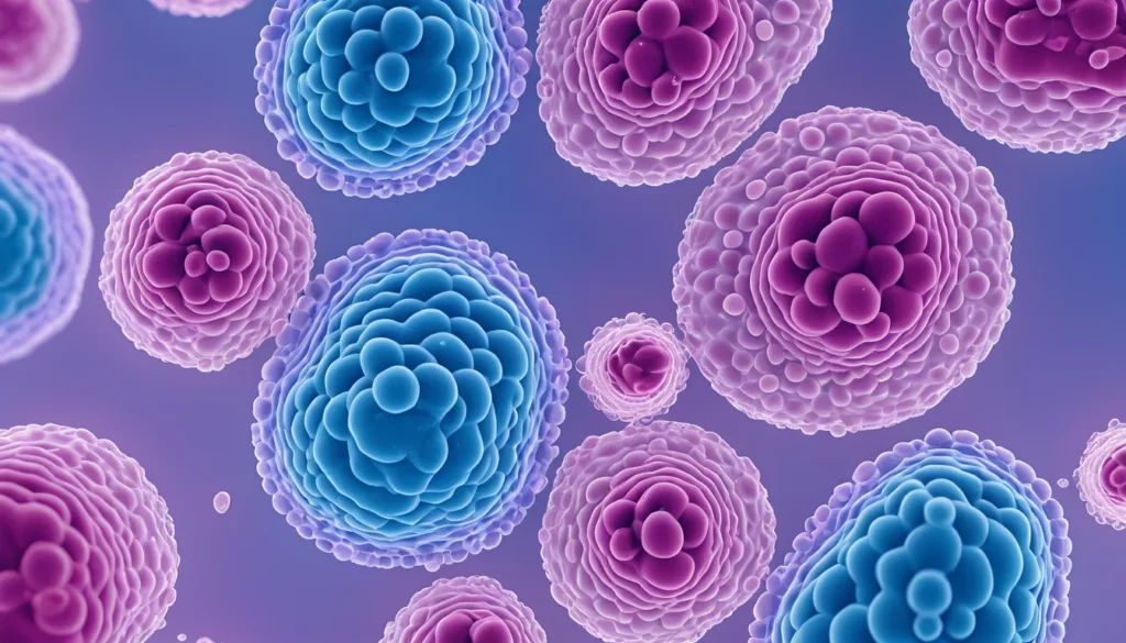
In practice, this means that acidophilic cells take up stains like eosin — a pink or red acidic dye — very easily. As a result, these cells appear bright pink or red when viewed under a microscope. This colour contrast helps pathologists identify different types of cells and tissues quickly and accurately.
The staining technique most commonly used is called haematoxylin and eosin (H&E) staining. It is one of the most widely used methods in diagnostic pathology around the world, including in Canadian hospital laboratories.
How Acidophilic Staining Works in the Lab
When a tissue sample is collected — for example, through a biopsy — it is sent to a pathology lab. There, technicians prepare thin slices of the tissue and apply a series of dyes. The haematoxylin dye stains acidic structures (like cell nuclei) blue or purple. The eosin dye stains basic or acidophilic structures pink or red.
Acidophilic cells absorb the eosin dye because they contain positively charged proteins and other molecules. These molecules attract the negatively charged eosin dye. Therefore, cells rich in proteins — such as muscle fibres and red blood cells — typically appear pink or red on the slide.
This process gives pathologists a detailed colour map of the tissue. They can then identify healthy cells, abnormal cells, and signs of disease. Learn more about how biopsy and tissue analysis works at Mayo Clinic.
Types of Staining Patterns
Not all cells stain the same way. Pathologists classify cells by how they react to dyes:
Acidophilic (eosinophilic): Cells that absorb acidic dyes and appear pink or red.
Basophilic: Cells that absorb basic dyes and appear blue or purple.
Neutrophilic: Cells that do not have a strong affinity for either type of dye and appear pale.
Understanding these patterns helps doctors distinguish between normal and abnormal tissue. Furthermore, certain diseases cause predictable changes in staining patterns, which makes the process a powerful diagnostic tool.
Where Acidophilic Cells Appear in the Body
Acidophilic cells are found throughout the human body. They are not limited to one organ or system. However, they are especially prominent in certain tissues that contain high concentrations of proteins.
Muscle Tissue
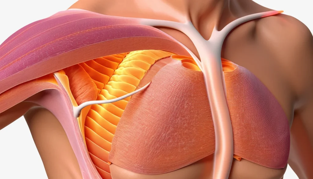
Muscle cells are strongly acidophilic. They contain large amounts of the proteins actin and myosin, which give them their pink colour under H&E staining. This makes it easier for pathologists to identify muscle tissue and spot any damage or disease, such as muscular dystrophy or inflammatory myopathy.
Red Blood Cells
Red blood cells (erythrocytes) are among the most recognisable acidophilic structures in histology. They are filled with haemoglobin, a protein that stains bright pink with eosin. Because of this, red blood cells are often used as a reference point when reading stained slides.
Liver Cells (Hepatocytes)
Liver cells are moderately acidophilic. In conditions like viral hepatitis or liver cell death (necrosis), hepatocytes can become intensely acidophilic. A deeply stained, shrunken liver cell is called an acidophilic body or Councilman body. This finding often signals significant liver injury.
Eosinophils
Eosinophils are a type of white blood cell named for their strong affinity for eosin stain. They play an important role in the immune system, particularly in fighting parasites and regulating allergic responses. Elevated levels of eosinophils in tissue can indicate allergic conditions, asthma, or certain infections.
Why Acidophilic Changes Matter for Your Health
When a pathologist reports acidophilic changes in a tissue sample, it is important to understand what that finding means in context. In many cases, it simply confirms the identity of normal, healthy tissue. However, in other cases, it signals cellular stress, injury, or disease.
For example, acidophilic change in liver cells is a hallmark of conditions like alcoholic liver disease, non-alcoholic fatty liver disease (NAFLD), and viral hepatitis. Similarly, acidophilic degeneration in kidney tubule cells may point to toxic injury or reduced blood supply.
In the stomach lining, certain cells called parietal cells are naturally acidophilic. Changes in the number or appearance of these cells can indicate conditions like gastritis or Helicobacter pylori infection. Healthline provides a helpful overview of gastritis and its causes.
Acidophilic Bodies as a Disease Marker
As mentioned earlier, acidophilic bodies are a specific pathological finding. They represent cells that have undergone a form of programmed cell death called apoptosis. Finding these bodies in tissue samples helps doctors confirm certain diagnoses.
In liver disease, acidophilic bodies strongly suggest hepatocyte apoptosis. In skin conditions like lichen planus or lupus, similar acidophilic bodies (called colloid or hyaline bodies) appear in the lower layers of the skin. These findings guide treatment decisions.
Acidophilic Cells and Cancer Diagnosis
Acidophilic staining also plays a role in identifying cancer cells. Many tumour cells show altered staining patterns compared to normal tissue. Some cancers produce cells that are strongly acidophilic, which can help pathologists classify the tumour type and grade.
For instance, certain types of kidney cancer (renal cell carcinoma), thyroid cancer (Hürthle cell tumours), and pituitary tumours contain cells with abundant acidophilic cytoplasm. Recognising these patterns is critical for accurate cancer staging and treatment planning.
In addition, changes in acidophilic staining can indicate how aggressive a tumour is. Therefore, pathology reports that mention acidophilic features are an important part of your overall cancer care. Health Canada offers resources on navigating the Canadian healthcare system for chronic and complex conditions.
Interpreting Your Pathology Report
If you receive a pathology report that mentions acidophilic cells or acidophilic change, do not panic. These terms describe what the tissue looks like under the microscope. They do not automatically mean something is seriously wrong.
Your doctor will interpret these findings alongside your symptoms, blood tests, and imaging results. Always ask your family doctor or specialist to walk you through your pathology report in plain language. Understanding your results is your right as a patient.
The Role of Histology in Canadian Healthcare
Histology — the study of tissue under a microscope — is a cornerstone of Canadian diagnostic medicine. Most provincial health plans cover biopsy and pathology services when ordered by a physician. This means that if your doctor suspects a problem, a tissue examination is accessible to you without direct out-of-pocket cost in most provinces.
Pathology labs across Canada follow strict quality standards. They ensure that staining procedures like H&E are performed consistently and accurately. As a result, whether you receive care in British Columbia or Newfoundland, the interpretation of acidophilic cells follows the same scientific principles.
Histological analysis is used across many medical specialties in Canada, including gastroenterology, oncology, dermatology, and nephrology. It is one of the most reliable tools available to your healthcare team.
How Samples Are Collected in Canada
Tissue samples are usually collected through a biopsy. This can happen during a procedure like a colonoscopy, endoscopy, or minor surgical excision. Your family doctor or a specialist will arrange this if needed. The sample is then sent to a hospital or community pathology lab for analysis.
Results typically take several days to two weeks. Once your doctor receives the report, they will contact you to discuss the findings and next steps. If you have concerns while waiting, do not hesitate to call your doctor’s office or visit a walk-in clinic for guidance.
When to See a Doctor
You do not need to seek medical care specifically about acidophilic cells — this is a laboratory term your doctor uses, not a symptom you would notice yourself. However, certain symptoms may prompt your doctor to order a biopsy or tissue analysis.
See your family doctor or visit a walk-in clinic if you experience any of the following:
Unexplained fatigue or weight loss
Persistent abdominal pain or changes in digestion
Yellowing of the skin or eyes (jaundice)
Unusual skin changes, lumps, or lesions
Blood in your urine or stool
Recurring allergic reactions or breathing difficulties
These symptoms may have many causes, most of which are manageable. However, early investigation — including tissue sampling when appropriate — leads to better outcomes. Your provincial health plan covers most diagnostic tests when referred by a physician.
If you have already received a pathology report mentioning acidophilic changes and you are unsure what it means, book an appointment with your doctor. They are the best person to explain what the findings mean for your specific situation.
Frequently Asked Questions
What does it mean when cells are acidophilic?
When cells are described as acidophilic, it means they absorb acidic dyes — like eosin — and appear pink or red under a microscope. This is a normal property of many cell types, including muscle cells and red blood cells. However, in some organs, acidophilic changes can indicate cell injury or disease.
Are acidophilic cells the same as eosinophilic cells?
Yes, the terms acidophilic and eosinophilic are often used interchangeably in histology. Both describe cells or structures that stain pink or red with eosin dye. The term eosinophilic is slightly more common in modern pathology reports, but they refer to the same staining behaviour.
What causes acidophilic change in liver cells?
Acidophilic change in liver cells is often caused by cell injury or death through a process called apoptosis. Common causes include viral hepatitis, alcoholic liver disease, and non-alcoholic fatty liver disease. A pathologist identifies these acidophilic cells in a biopsy sample to help diagnose the underlying condition.
Is an acidophilic body a sign of cancer?
An acidophilic body is not always a sign of cancer. It most commonly indicates apoptosis, or programmed cell death, which can occur in inflammatory conditions, infections, and liver disease. However, your doctor will always consider the full picture — including your symptoms and other test results — before drawing any conclusions.
Why do pathologists use eosin stain to identify acidophilic cells?
Pathologists use eosin stain because it binds strongly to the positively charged proteins inside acidophilic cells, producing a bright pink or red colour that is easy to see under a microscope. This colour contrast makes it simple to distinguish different tissue types and spot abnormalities. The haematoxylin and eosin (H&E) method has been a standard tool in diagnostic pathology for over a century.
Can I request my pathology results in Canada?
Yes, in Canada you have the right to access your own medical records, including pathology reports. You can request these through your family doctor, the hospital where the procedure was performed, or your provincial health authority. It is always a good idea to ask your doctor to explain the meaning of terms like acidophilic cells so you fully understand your results.
Key Takeaways
Acidophilic cells are cells that absorb acidic dyes and appear pink or red under a microscope during tissue staining.
The most common staining method used in Canadian labs is haematoxylin and eosin (H&E) staining.
Many healthy cell types — including muscle cells and red blood cells — are naturally acidophilic.
Acidophilic changes in organ cells, such as liver cells, can signal injury, inflammation, or disease.
Acidophilic bodies are a specific finding linked to apoptosis and are seen in conditions like hepatitis and certain skin disorders.
Pathology and biopsy services are covered under most provincial health plans in Canada.
If you receive a pathology report mentioning acidophilic findings, speak with your family doctor or specialist to understand what it means for your health.
This article is intended for general health information only. Always consult your family doctor, walk-in clinic physician, or a qualified medical specialist for advice about your personal health situation.