The acanthion is a small but important point on the human skull. It sits at the tip of the anterior nasal spine — the tiny bony projection at the base of the nose. Medical professionals use this craniometric landmark in facial analysis, surgical planning, and diagnostic imaging. Understanding the acanthion can help you make sense of what your doctor or specialist might be measuring during a facial or skull assessment.
What Is the Acanthion?
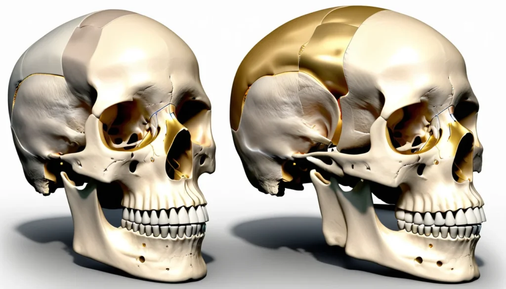
The acanthion is one of many craniometric points on the human skull. A craniometric point is simply a fixed, identifiable spot on the skull that doctors and scientists use as a reference. Think of these points as landmarks on a map — they help professionals navigate the complex geography of the human face and head.
Specifically, the acanthion sits at the very tip of the anterior nasal spine. The anterior nasal spine is a small, pointed projection of bone located at the bottom of the nasal opening. You can feel this general area if you gently press just above your upper lip, at the base of your nose.
This point plays a key role in many areas of medicine. For example, surgeons, orthodontists, radiologists, and forensic scientists all rely on the acanthion when they need precise facial measurements.
Why the Acanthion Matters in Medicine
Craniometric points like the acanthion are essential tools in modern medicine. They give healthcare providers a consistent, reproducible way to measure and compare facial structures. Without these fixed reference points, accurate facial analysis would be nearly impossible.
Furthermore, the acanthion is used in a variety of medical fields. Each discipline uses it slightly differently, but the goal is always the same — precision and consistency in measurement.
Orthodontics and Dentistry
Orthodontists frequently use the acanthion when analysing X-rays of the skull, called cephalometric radiographs. These X-rays show the bones of the skull and jaw in profile. By measuring distances between the acanthion and other craniometric points, orthodontists can assess how the jaw and teeth align. This helps them plan treatments such as braces or corrective jaw surgery.
In Canada, your orthodontist or dentist may take these types of X-rays during a detailed assessment. Provincial health plans may cover some of these diagnostic imaging costs, particularly for children. Check with your provincial health authority or family doctor to understand your coverage.
Maxillofacial Surgery
Maxillofacial surgeons — specialists who operate on the face, mouth, and jaw — rely heavily on craniometric landmarks. The acanthion serves as a reference point when surgeons plan operations to correct structural issues with the nose, jaw, or midface region. Accurate measurements help ensure the best possible outcomes for patients.
Therefore, if you are ever referred to a maxillofacial surgeon, do not be surprised if they take detailed measurements of your facial structure. These measurements are a standard part of surgical planning.
Radiology and Diagnostic Imaging
Radiologists use the acanthion when interpreting CT scans and MRI images of the face and skull. It helps them identify and describe the exact location of fractures, tumours, or other abnormalities. In addition, it allows different radiologists to communicate clearly with one another using shared reference points.
Many Canadian hospitals and imaging centres use standardised craniometric terminology. This ensures that a radiologist in Vancouver and one in Halifax are both describing the same thing when they refer to the acanthion.
The Acanthion in Forensic Science
Forensic scientists use craniometric points, including the acanthion, to help identify human remains. When a skull is recovered, forensic anthropologists take a series of measurements between key landmarks. These measurements can help determine a person’s age, sex, and ancestry. In some cases, they can even help reconstruct what a person looked like.
This work is crucial in criminal investigations, mass disaster identification, and historical research. The Royal Canadian Mounted Police (RCMP) and Canadian forensic teams use these same scientific principles in their investigative work.
However, forensic craniometry is a highly specialised field. It requires years of training and should only be performed by qualified professionals. The acanthion is just one of dozens of landmarks used in this process.
The Acanthion in Anthropology and Human Evolution
Beyond medicine and forensics, the acanthion has long been studied in physical anthropology. Researchers use craniometric data — including measurements involving the acanthion — to understand how human populations have changed over time. These studies help trace migration patterns, evolutionary changes, and population diversity across the globe.
For example, researchers have found that the shape and position of the anterior nasal spine, where the acanthion is located, can vary between different human populations. These variations are studied carefully and respectfully as part of understanding human biological diversity.
As a result, the acanthion is not just a medical term — it is also a window into human history and evolution. The World Health Organization provides resources on human health research and global health standards that inform many of these scientific studies.
How the Acanthion Is Measured
Measuring the acanthion is a precise process. Healthcare professionals typically identify it using one of two methods: direct measurement on a patient, or measurement from medical imaging such as X-rays, CT scans, or MRI scans.
Direct Measurement
In a clinical setting, a trained professional locates the anterior nasal spine by touch and marks or identifies the acanthion as its most forward-projecting point. They then use calibrated instruments to measure the distance between the acanthion and other craniometric landmarks. This type of measurement is common in orthodontic and anthropological assessments.
Imaging-Based Measurement
In radiology and surgery, the acanthion is identified on digital images. Software tools allow clinicians to mark the point precisely and calculate distances automatically. This method is extremely accurate and is the standard approach in surgical planning and research settings.
In addition, digital craniometry has become more common across Canadian healthcare settings as hospitals adopt advanced imaging technology. Mayo Clinic offers detailed information on how imaging is used in facial and skull assessments, which may help you understand what to expect during a specialist appointment.
Common Conditions That Involve the Anterior Nasal Spine Area
While the acanthion itself is simply a reference point, the bone it sits on — the anterior nasal spine — can be affected by several medical conditions. Understanding these conditions can help you recognise when to seek medical advice.
Nasal Fractures
A blow to the nose can fracture the anterior nasal spine. This type of fracture may not always be obvious on the surface. However, it can cause pain, swelling, and difficulty breathing through the nose. If you have taken a hard hit to the nose, it is important to seek medical attention promptly.
Nasal Deviation and Septal Issues
A deviated nasal septum — when the wall between your nostrils is off-centre — can sometimes affect the position of the anterior nasal spine. This may contribute to breathing difficulties or chronic nasal congestion. Surgeons use the acanthion as a reference point when correcting these structural issues.
Congenital Facial Differences
Some people are born with structural differences in the midface region, such as cleft lip and palate. These conditions can affect the shape and position of the anterior nasal spine and the acanthion. Craniometric measurements, including those involving the acanthion, help surgeons plan corrective procedures for these conditions.
In Canada, children with cleft lip and palate receive multidisciplinary care through specialised teams at children’s hospitals across the country. Provincial health plans typically cover these treatments. Health Canada provides information on health services and coverage available to Canadians.
When to See a Doctor
Most Canadians will never need to think about the acanthion in their daily lives. However, there are situations where a healthcare provider may assess this area as part of a broader evaluation. Knowing when to seek care is always important.
You should see your family doctor or visit a walk-in clinic if you experience any of the following:
Pain or swelling at the base of your nose after an injury
Difficulty breathing through your nose that does not improve
Visible changes in the shape of your nose after a trauma
Chronic nasal congestion that affects your quality of life
Any concerns about facial asymmetry or structural changes in your face
Your family doctor can assess your symptoms and refer you to the appropriate specialist, such as an ear, nose, and throat (ENT) doctor or a maxillofacial surgeon, if needed. If you do not have a family doctor, a walk-in clinic is a great starting point for getting care in most Canadian provinces and territories.
Always consult a qualified healthcare professional before drawing any conclusions about your health based on medical terminology you have read online. This article is for general information only and is not a substitute for professional medical advice.
Frequently Asked Questions About the Acanthion
What is the acanthion in anatomy?
The acanthion is a craniometric point located at the tip of the anterior nasal spine, a small bony projection at the base of the nose. Healthcare professionals use the acanthion as a fixed reference point when measuring and analysing facial structures. It is used in fields such as orthodontics, surgery, radiology, and forensic science.
Where exactly is the acanthion located on the skull?
The acanthion sits at the most forward-projecting tip of the anterior nasal spine, which is the pointed bony ridge at the very base of the nasal opening. You can roughly locate this area by gently pressing just above your upper lip at the centre of your nose. It is a standard landmark used in cephalometric X-rays and facial assessments.
Why do doctors and surgeons use craniometric points like the acanthion?
Doctors and surgeons use craniometric points, including the acanthion, because they provide consistent and reproducible reference locations on the skull. These landmarks allow healthcare professionals to take precise measurements, plan surgeries, and compare results across patients. Without these fixed points, accurate facial analysis and surgical planning would be far more difficult.
Is the acanthion relevant to orthodontic treatment?
Yes, the acanthion is commonly used in orthodontic cephalometric analysis. Orthodontists measure the distance between the acanthion and other skull landmarks on profile X-rays to assess how the jaw and teeth relate to the rest of the face. These measurements help guide treatment planning for braces, retainers, and corrective jaw surgery.
Can the anterior nasal spine, where the acanthion is located, be fractured?
Yes, the anterior nasal spine can be fractured following a direct blow to the nose or midface area. A fracture in this region may cause pain, swelling, and nasal breathing difficulties. If you suspect a nasal fracture, you should visit a walk-in clinic or emergency department for a proper assessment.
How is the acanthion used in forensic science?
In forensic science, the acanthion is one of many craniometric landmarks used to help identify human remains. Forensic anthropologists measure distances between skull landmarks to estimate a person’s age, sex, and ancestry. The acanthion is a key reference point in this process because of its consistent and easily identifiable location on the skull.
Key Takeaways
The acanthion is a craniometric point located at the tip of the anterior nasal spine, at the base of the nose.
It serves as a precise reference point used in orthodontics, maxillofacial surgery, radiology, forensic science, and anthropology.
Healthcare professionals use the acanthion to take accurate facial measurements and plan medical or surgical treatments.
The anterior nasal spine, where the acanthion is located, can be affected by fractures, septal deviation, and congenital conditions.
If you have concerns about nasal pain, breathing difficulties, or facial structure, speak with your family doctor or visit a walk-in clinic.
This article is for informational purposes only. Always consult a qualified healthcare professional for personal medical advice.